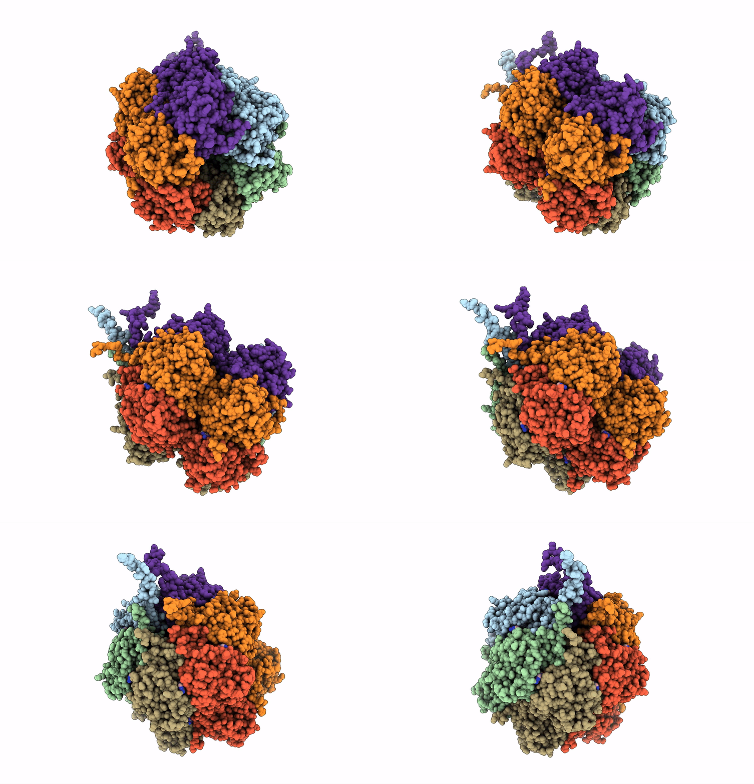
2GBL image
Deposition Date 2006-03-10
Release Date 2007-01-23
Last Version Date 2024-10-09
Entry Detail
PDB ID:
2GBL
Title:
Crystal Structure of Full Length Circadian Clock Protein KaiC with Phosphorylation Sites
Biological Source:
Source Organism:
Method Details:
Experimental Method:
Resolution:
2.80 Å
R-Value Free:
0.29
R-Value Work:
0.23
R-Value Observed:
0.23
Space Group:
P 21 21 21
Macromolecular Entities
Structures with similar UniProt ID
Protein Blast
Polymer Type:polypeptide(L)
Molecule:Circadian clock protein kinase kaiC
Gene (Uniprot):kaiC
Chain IDs:A, B, E, F
Chain Length:519
Number of Molecules:4
Biological Source:Synechococcus elongatus
Structures with similar UniProt ID
Protein Blast
Polymer Type:polypeptide(L)
Molecule:Circadian clock protein kinase kaiC
Gene (Uniprot):kaiC
Chain IDs:C, D
Chain Length:519
Number of Molecules:2
Biological Source:Synechococcus elongatus
Modified Residue
Compound ID Chain ID Parent Comp ID Details 2D Image
SEP A SER PHOSPHOSERINE
TPO A THR PHOSPHOTHREONINE
Primary Citation
Analysis of KaiA-KaiC protein interactions in the cyano-bacterial circadian clock using hybrid structural methods.
Embo J. 25 2017 2028 (2006)
PMID: 16628225 DOI: 10.1038/sj.emboj.7601086

Abstact

The cyanobacterial circadian clock can be reconstituted in vitro by mixing recombinant KaiA, KaiB and KaiC proteins with ATP, producing KaiC phosphorylation and dephosphorylation cycles that have a regular rhythm with a ca. 24-h period and are temperature-compensated. KaiA and KaiB are modulators of KaiC phosphorylation, whereby KaiB antagonizes KaiA's action. Here, we present a complete crystallographic model of the Synechococcus elongatus KaiC hexamer that includes previously unresolved portions of the C-terminal regions, and a negative-stain electron microscopy study of S. elongatus and Thermosynechococcus elongatus BP-1 KaiA-KaiC complexes. Site-directed mutagenesis in combination with EM reveals that KaiA binds exclusively to the CII half of the KaiC hexamer. The EM-based model of the KaiA-KaiC complex reveals protein-protein interactions at two sites: the known interaction of the flexible C-terminal KaiC peptide with KaiA, and a second postulated interaction between the apical region of KaiA and the ATP binding cleft on KaiC. This model brings KaiA mutation sites that alter clock period or abolish rhythmicity into contact with KaiC and suggests how KaiA might regulate KaiC phosphorylation.

Legend

Protein

Chemical

Disease

Primary Citation of related structures