2GBK image
Deposition Date 2006-03-10
Release Date 2006-05-16
Last Version Date 2023-08-30
Entry Detail
PDB ID:
2GBK
Keywords:
Title:
Crystal Structure of the 9-10 MoaD Insertion Mutant of Ubiquitin
Biological Source:
Source Organism:
Homo sapiens (Taxon ID: 9606)
Method Details:
Experimental Method:
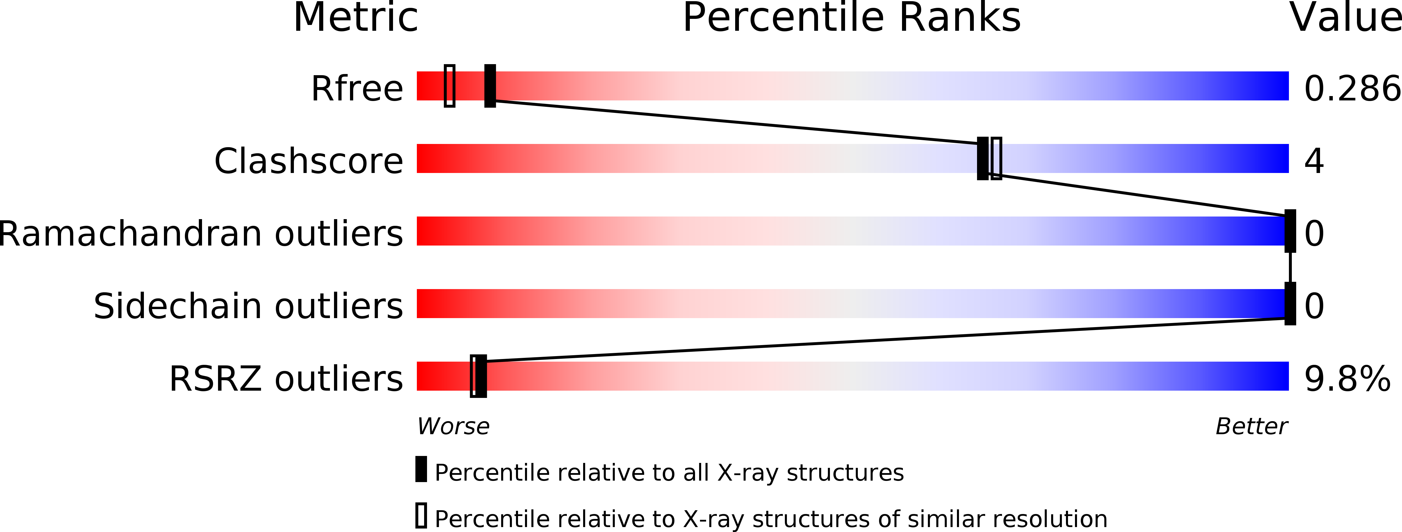
Resolution:
1.99 Å
R-Value Free:
0.28
R-Value Work:
0.24
R-Value Observed:
0.24
Space Group:
P 1
Macromolecular Entities
Structures with similar UniProt ID
Protein Blast
Polymer Type:polypeptide(L)
Molecule:Ubiquitin
Chain IDs:A, B, C, D
Chain Length:83
Number of Molecules:4
Biological Source:Homo sapiens
Primary Citation
Structures of Ubiquitin Insertion Mutants Support Site-specific Reflex Response to Insertions Hypothesis.
J.Mol.Biol. 359 390 402 (2006)
PMID: 16647719 DOI: 10.1016/j.jmb.2006.03.047

Abstact

We previously concluded that, judging from NMR chemical shifts, the effects of insertions into ubiquitin on its conformation appear to depend primarily on the site of insertion rather than the sequence of the insertion. To obtain a more complete and atomic-resolution understanding of how these insertions modulate the conformation of ubiquitin, we have solved the crystal structures of four insertional mutants of ubiquitin. Insertions between residues 9 and 10 of ubiquitin are minimally perturbing to the remainder of the protein, while larger alterations occur when the insertion is between residues 35 and 36. Further, the alterations in response to insertions are very similar for each mutant at a given site. Two insertions, one at each site, were designed from structurally homologous proteins. Interestingly, the secondary structure within these five to seven amino acid residue insertions is conserved in the new protein. Overall, the crystal structures support the previous conclusion that the conformational effects of these insertions are determined largely by the site of insertion and only secondarily by the sequence of the insert.

Legend

Protein

Chemical

Disease

Primary Citation of related structures