2GB0 image
Deposition Date 2006-03-09
Release Date 2006-03-21
Last Version Date 2024-10-09
Entry Detail
PDB ID:
2GB0
Keywords:
Title:
Monomeric sarcosine oxidase: structure of a covalently flavinylated amine oxidizing enzyme
Biological Source:
Source Organism:
Bacillus sp. (Taxon ID: 69000)
Host Organism:
Method Details:
Experimental Method:
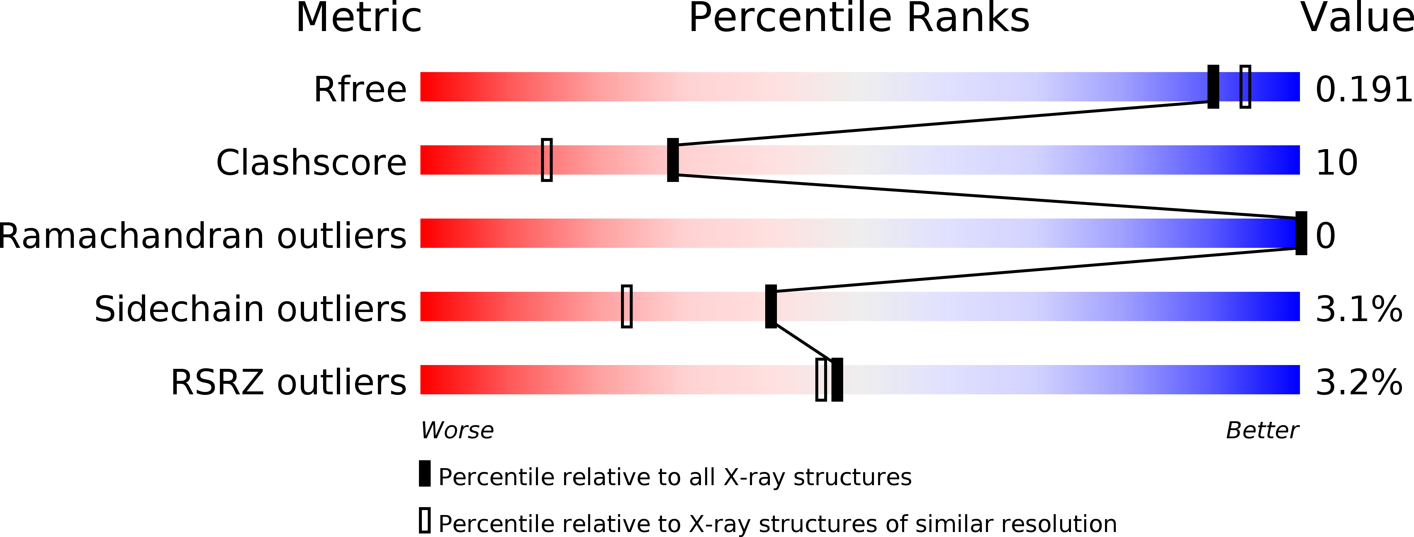
Resolution:
1.85 Å
R-Value Free:
0.19
R-Value Work:
0.16
Space Group:
P 1 21 1
Macromolecular Entities
Structures with similar UniProt ID
Protein Blast
Polymer Type:polypeptide(L)
Molecule:Monomeric sarcosine oxidase
Gene (Uniprot):soxA
Chain IDs:A, B
Chain Length:389
Number of Molecules:2
Biological Source:Bacillus sp.
Primary Citation
Monomeric Sarcosine Oxidase: Structure of a Covalently Flavinylated Amine Oxidizing Enzyme
Structure 7 331 345 (1999)
PMID: 10368302 DOI: 10.1016/S0969-2126(99)80043-4

Abstact

BACKGROUND: Monomeric sarcosine oxidases (MSOXs) are among the simplest members of a recently recognized family of eukaryotic and prokaryotic enzymes that catalyze similar oxidative reactions with various secondary or tertiary amino acids and contain covalently bound flavins. Other members of this family include heterotetrameric sarcosine oxidase, N-methyltryptophan oxidase and pipecolate oxidase. Mammalian sarcosine dehydrogenase and dimethylglycine dehydrogenase may be more distantly related family members. RESULTS: The X-ray crystal structure of MSOX from Bacillus sp. B-0618, expressed in Escherichia coli, has been solved at 2.0 A resolution by multiwavelength anomalous dispersion (MAD) from crystals of the selenomethionine-substituted enzyme. Fourteen selenium sites, belonging to two MSOX molecules in the asymmetric unit, were used for MAD phasing and to define the local twofold symmetry axis for electron-density averaging. The structures of the native enzyme and of two enzyme-inhibitor complexes were also determined. CONCLUSIONS: MSOX is a two-domain protein with an overall topology most similar to that of D-amino acid oxidase, with which it shares 14% sequence identity. The flavin ring is located in a very basic environment, making contact with sidechains of arginine, lysine, histidine and the N-terminal end of a helix dipole. The flavin is covalently attached through an 8alpha-S-cysteinyl linkage to Cys315 of the catalytic domain. Covalent attachment is probably self-catalyzed through interactions with the positive sidechains and the helix dipole. Substrate binding is probably stabilized by hydrogen bonds between the substrate carboxylate and two basic sidechains, Arg52 and Lys348, located above the re face of the flavin ring.

Legend

Protein

Chemical

Disease

Primary Citation of related structures
Feedback Form
Name
Email
Institute
Feedback