
Deposition Date
2006-03-08
Release Date
2006-08-08
Last Version Date
2024-10-30
Entry Detail
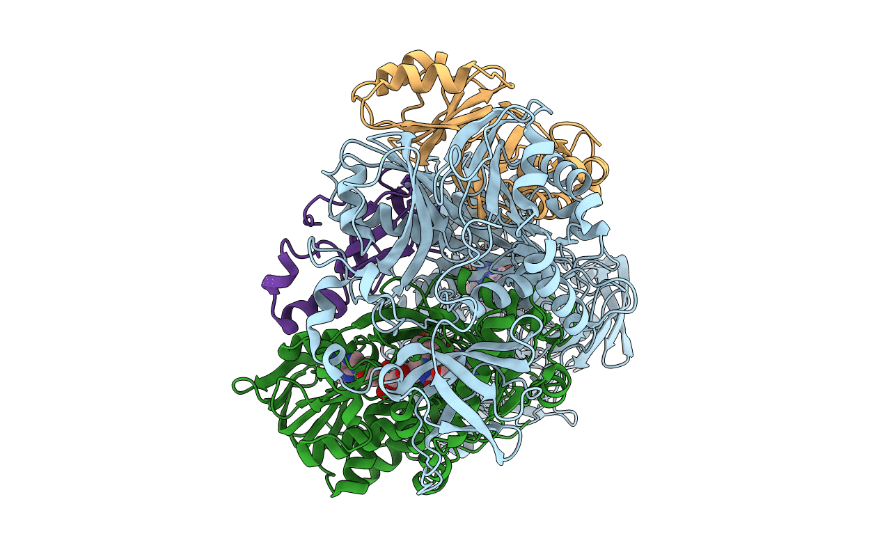
PDB ID:
2GAH
Keywords:
Title:
Heterotetrameric sarcosine: structure of a diflavin metaloenzyme at 1.85 a resolution
Biological Source:
Source Organism(s):
Stenotrophomonas maltophilia (Taxon ID: 40324)
Expression System(s):
Method Details:
Experimental Method:
Resolution:
2.00 Å
R-Value Free:
0.21
R-Value Work:
0.17
Space Group:
P 21 21 21
