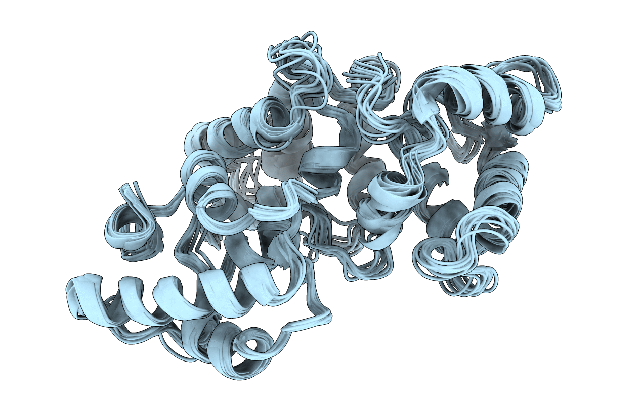
2G9B image
Entry Detail
PDB ID:
2G9B
Title:
NMR solution structure of CA2+-loaded calbindin D28K
Biological Source:
Source Organism:
Host Organism:
PDB Version:
Deposition Date:
2006-03-06
Release Date:
2006-07-04
Method Details:
Experimental Method:
Conformers Calculated:
900
Conformers Submitted:
10
Selection Criteria:
structures with the least restraint violations, structures with the lowest restraint energy
Macromolecular Entities
Polymer Type:polypeptide(L)
Description:Calbindin
Chain IDs:A
Chain Length:263
Number of Molecules:1
Biological Source:Rattus norvegicus
Ligand Molecules
Primary Citation
Structure, binding interface and hydrophobic transitions of Ca(2+)-loaded calbindin-D(28K).
Nat.Struct.Mol.Biol. 13 641 647 (2006)
PMID: 16799559 DOI: 10.1038/nsmb1112

Abstact

Calbindin-D(28K) is a Ca2+-binding protein, performing roles as both a calcium buffer and calcium sensor. The NMR solution structure of Ca2+-loaded calbindin-D(28K) reveals a single, globular fold consisting of six distinct EF-hand subdomains, which coordinate Ca2+ in loops on EF1, EF3, EF4 and EF5. Target peptides from Ran-binding protein M and myo-inositol monophosphatase, along with a new target from procaspase-3, are shown to interact with the protein on a surface comprised of alpha5 (EF3), alpha8 (EF4) and the EF2-EF3 and EF4-EF5 loops. Fluorescence experiments reveal that calbindin-D(28K) adopts discrete hydrophobic states as it binds Ca2+. The structure, binding interface and hydrophobic characteristics of Ca2+-loaded calbindin-D(28K) provide the first detailed insights into how this essential protein may function. This structure is one of the largest high-resolution NMR structures and the largest monomeric EF-hand protein to be solved to date.

Legend

Protein

Chemical

Disease

Primary Citation of related structures