2G7I image
Deposition Date 2006-02-28
Release Date 2006-05-23
Last Version Date 2024-10-30
Entry Detail
PDB ID:
2G7I
Keywords:
Title:
Structure of Human Complement Factor H Carboxyl Terminal Domains 19-20: a Basis for Atypical Hemolytic Uremic Syndrome
Biological Source:
Source Organism:
Homo sapiens (Taxon ID: 9606)
Host Organism:
Method Details:
Experimental Method:
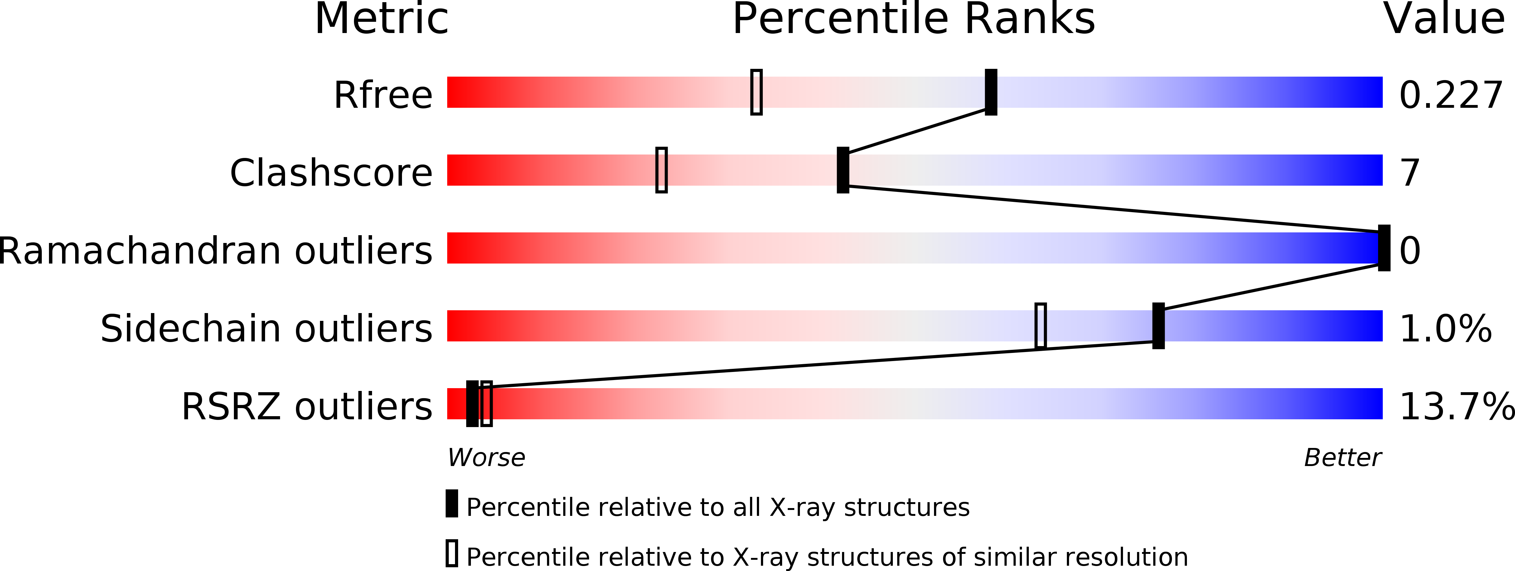
Resolution:
1.75 Å
R-Value Free:
0.22
R-Value Work:
0.20
R-Value Observed:
0.20
Space Group:
I 41 2 2
Macromolecular Entities
Structures with similar UniProt ID
Protein Blast
Polymer Type:polypeptide(L)
Molecule:Complement factor H
Gene (Uniprot):CFH
Chain IDs:A
Chain Length:125
Number of Molecules:1
Biological Source:Homo sapiens
Primary Citation
Structure of complement factor H carboxyl-terminus reveals molecular basis of atypical haemolytic uremic syndrome.
Embo J. 25 1784 1794 (2006)
PMID: 16601698 DOI: 10.1038/sj.emboj.7601052

Abstact

Factor H (FH) is the key regulator of the alternative pathway of complement. The carboxyl-terminal domains 19-20 of FH interact with the major opsonin C3b, glycosaminoglycans, and endothelial cells. Mutations within this area are associated with atypical haemolytic uremic syndrome (aHUS), a disease characterized by damage to endothelial cells, erythrocytes, and kidney glomeruli. The structure of recombinant FH19-20, solved at 1.8 A by X-ray crystallography, reveals that the short consensus repeat domain 20 contains, unusually, a short alpha-helix, and a patch of basic residues at its base. Most aHUS-associated mutations either destabilize the structure or cluster in a unique region on the surface of FH20. This region is close to, but distinct from, the primary heparin-binding patch of basic residues. By mutating five residues in this region, we show that it is involved, not in heparin, but in C3b binding. Therefore, the majority of the aHUS-associated mutations on the surface of FH19-20 interfere with the interaction between FH and C3b. This obviously leads to impaired control of complement attack on plasma-exposed cell surfaces in aHUS.

Legend

Protein

Chemical

Disease

Primary Citation of related structures