Deposition Date
2006-02-28
Release Date
2006-10-31
Last Version Date
2024-10-30
Entry Detail
PDB ID:
2G7E
Keywords:
Title:
The 1.6 A crystal structure of Vibrio cholerae extracellular endonuclease I
Biological Source:
Source Organism(s):
Vibrio cholerae (Taxon ID: 666)
Expression System(s):
Method Details:
Experimental Method:
Resolution:
1.60 Å
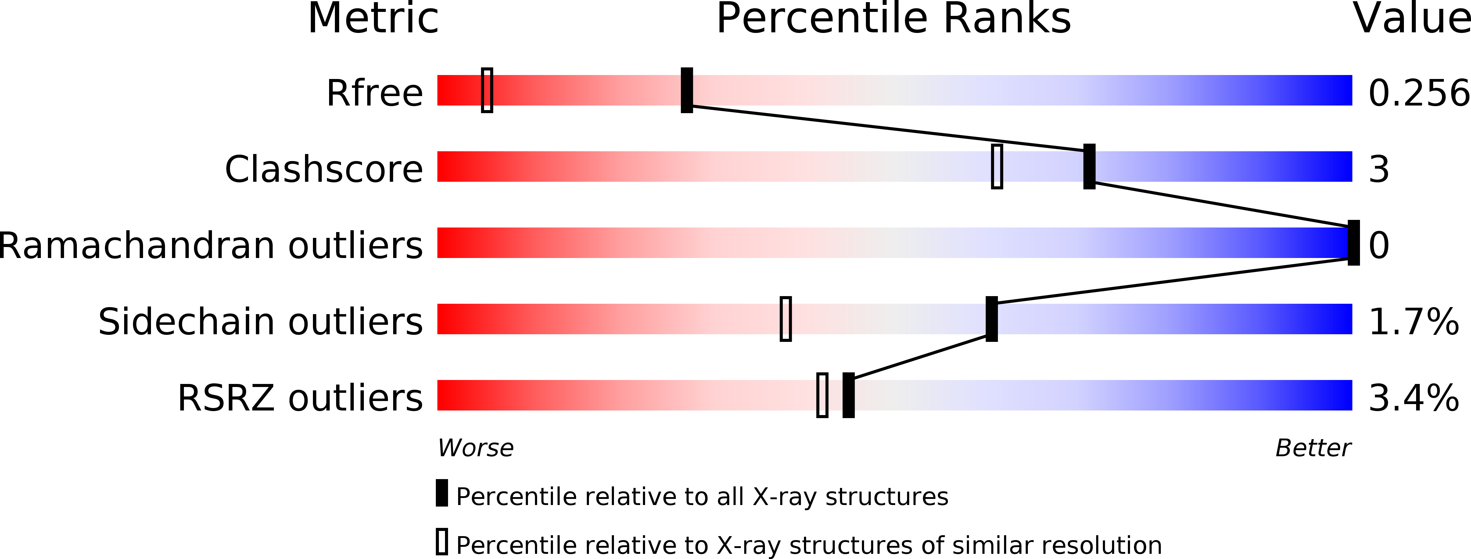
R-Value Free:
0.25
R-Value Work:
0.22
R-Value Observed:
0.22
Space Group:
P 21 21 21