2G6Y image
Deposition Date 2006-02-26
Release Date 2006-03-28
Last Version Date 2024-10-30
Entry Detail
PDB ID:
2G6Y
Title:
Crystal structure of the novel green fluorescent protein from marine copepod Pontellina plumata
Biological Source:
Source Organism:
Host Organism:
Method Details:
Experimental Method:
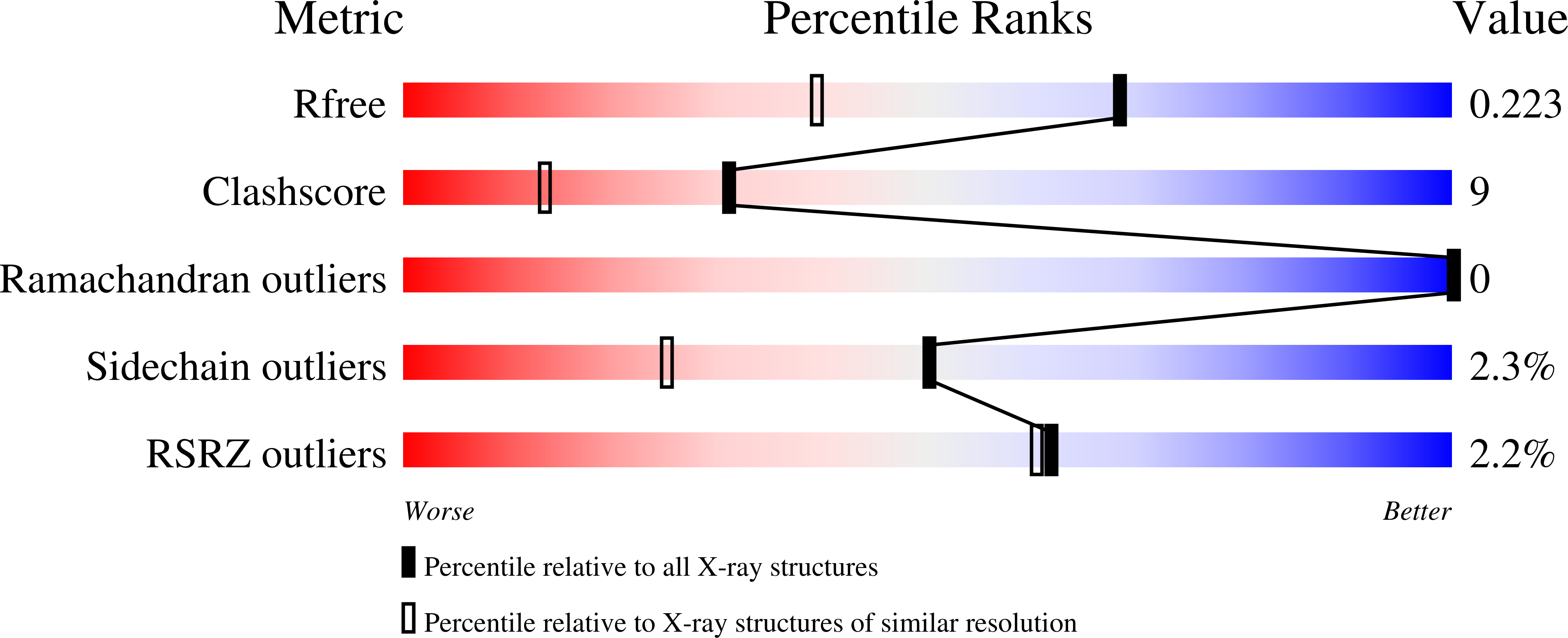
Resolution:
1.60 Å
R-Value Free:
0.21
R-Value Work:
0.16
R-Value Observed:
0.16
Space Group:
P 1 21 1
Macromolecular Entities
Polymer Type:polypeptide(L)
Molecule:green fluorescent protein 2
Mutations:K5E, V117M, V149D, V151D, A155T, F168S, L200D, I219D
Chain IDs:A, B, C, D
Chain Length:219
Number of Molecules:4
Biological Source:Pontellina plumata
Modified Residue
Compound ID Chain ID Parent Comp ID Details 2D Image
CR2 A GLY ?
Primary Citation
Structural basis for the fast maturation of Arthropoda green fluorescent protein.
Embo Rep. 7 1006 1012 (2006)
PMID: 16936637 DOI: 10.1038/sj.embor.7400787

Abstact

Since the cloning of Aequorea victoria green fluorescent protein (GFP) in 1992, a family of known GFP-like proteins has been growing rapidly. Today, it includes more than a hundred proteins with different spectral characteristics cloned from Cnidaria species. For some of these proteins, crystal structures have been solved, showing diversity in chromophore modifications and conformational states. However, we are still far from a complete understanding of the origin, functions and evolution of the GFP family. Novel proteins of the family were recently cloned from evolutionarily distant marine Copepoda species, phylum Arthropoda, demonstrating an extremely rapid generation of fluorescent signal. Here, we have generated a non-aggregating mutant of Copepoda fluorescent protein and solved its high-resolution crystal structure. It was found that the protein beta-barrel contains a pore, leading to the chromophore. Using site-directed mutagenesis, we showed that this feature is critical for the fast maturation of the chromophore.

Legend

Protein

Chemical

Disease

Primary Citation of related structures