2G4B image
Deposition Date 2006-02-21
Release Date 2006-07-25
Last Version Date 2023-08-30
Entry Detail
PDB ID:
2G4B
Title:
Structure of U2AF65 variant with polyuridine tract
Biological Source:
Source Organism:
Homo sapiens (Taxon ID: 9606)
Host Organism:
Method Details:
Experimental Method:
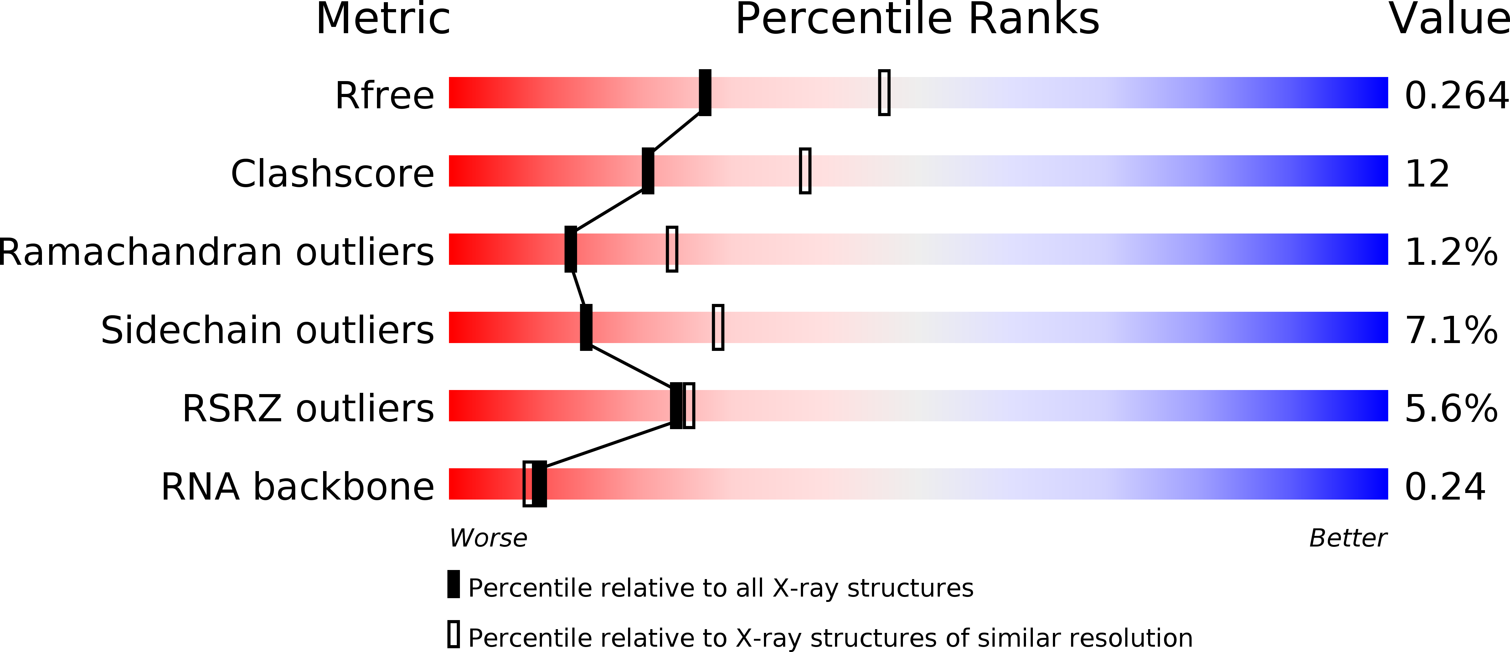
Resolution:
2.50 Å
R-Value Free:
0.27
R-Value Work:
0.26
R-Value Observed:
0.26
Space Group:
P 65 2 2
Macromolecular Entities
Polymer Type:polypeptide(L)
Molecule:Splicing factor U2AF 65 kDa subunit
Gene (Uniprot):U2AF2
Mutations:deletion of residues 238-258
Chain IDs:B (auth: A)
Chain Length:172
Number of Molecules:1
Biological Source:Homo sapiens
Ligand Molecules
Primary Citation
Structural basis of polypyrimidine tract recognition by the essential splicing factor U2AF65.
Mol.Cell 23 49 59 (2006)
PMID: 16818232 DOI: 10.1016/j.molcel.2006.05.025

Abstact

The essential pre-mRNA splicing factor, U2AF(65), guides the early stages of splice site choice by recognizing a polypyrimidine (Py) tract consensus sequence near the 3' splice site. Since Py tracts are relatively poorly conserved in higher eukaryotes, U2AF(65) is faced with the problem of specifying uridine-rich sequences, yet tolerating a variety of nucleotide substitutions found in natural Py tracts. To better understand these apparently contradictory RNA binding characteristics, the X-ray structure of the U2AF(65) RNA binding domain bound to a Py tract composed of seven uridines has been determined at 2.5 A resolution. Specific hydrogen bonds between U2AF(65) and the uracil bases provide an explanation for polyuridine recognition. Flexible side chains and bound water molecules form the majority of the base contacts and potentially could rearrange when the U2AF(65) structure adapts to different Py tract sequences. The energetic importance of conserved residues for Py tract binding is established by analysis of site-directed mutant U2AF(65) proteins using surface plasmon resonance.

Legend

Protein

Chemical

Disease

Primary Citation of related structures