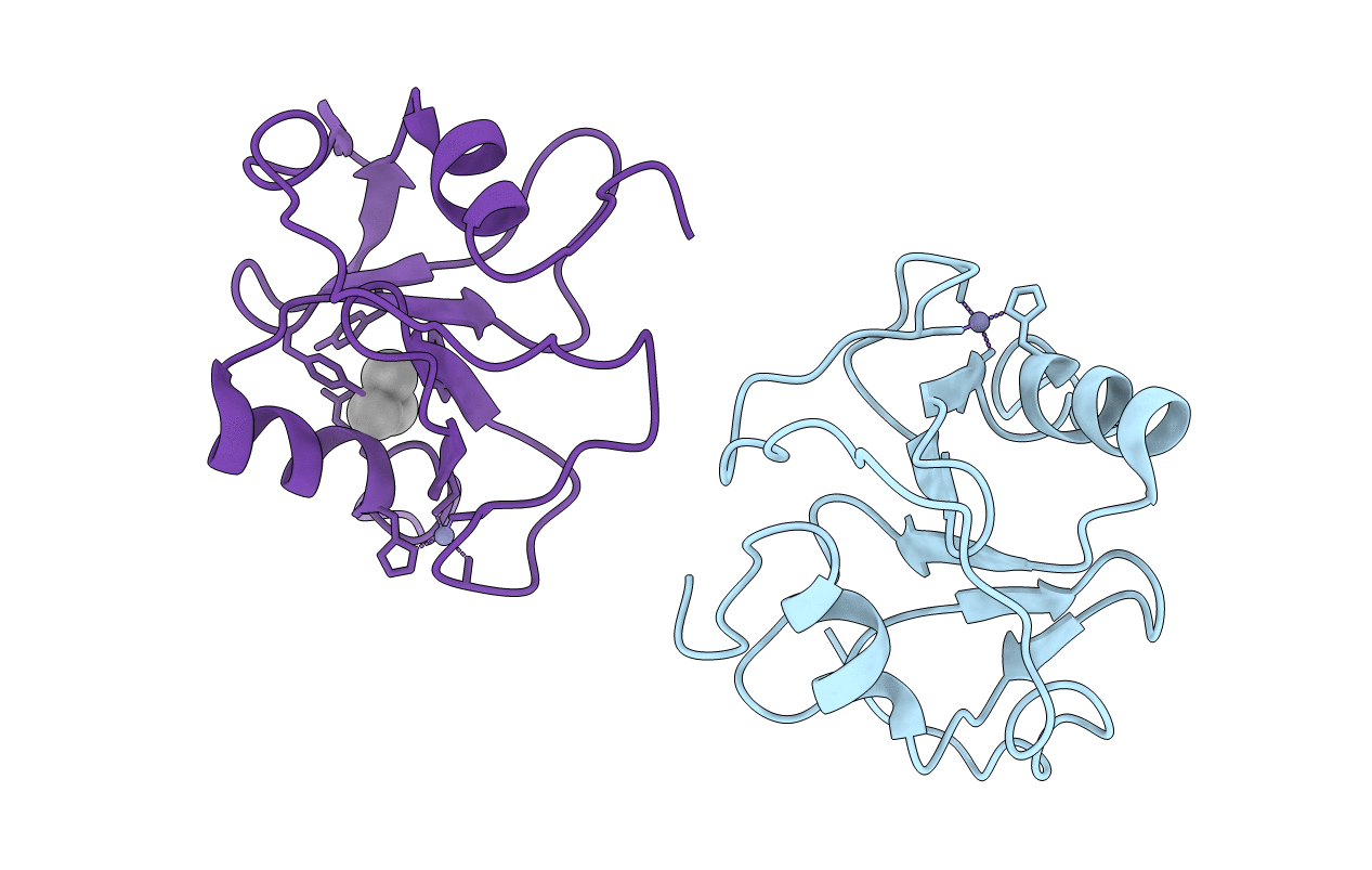
2G43 image
Entry Detail
PDB ID:
2G43
Keywords:
Title:
Structure of the ZNF UBP domain from deubiquitinating enzyme isopeptidase T (IsoT)
Biological Source:
Source Organism:
Host Organism:
PDB Version:
Deposition Date:
2006-02-21
Release Date:
2006-04-04
Method Details:
Experimental Method:
Resolution:
2.09 Å
R-Value Free:
0.27
R-Value Work:
0.22
R-Value Observed:
0.22
Space Group:
C 1 2 1
Macromolecular Entities
Polymer Type:polypeptide(L)
Description:Ubiquitin carboxyl-terminal hydrolase 5
Chain IDs:A, B
Chain Length:129
Number of Molecules:2
Biological Source:Homo sapiens
Primary Citation
The Ubiquitin Binding Domain ZnF UBP Recognizes the C-Terminal Diglycine Motif of Unanchored Ubiquitin.
Cell(Cambridge,Mass.) 124 1197 1208 (2006)
PMID: 16564012 DOI: 10.1016/j.cell.2006.02.038

Abstact

Ubiquitin binding proteins regulate the stability, function, and/or localization of ubiquitinated proteins. Here we report the crystal structures of the zinc-finger ubiquitin binding domain (ZnF UBP) from the deubiquitinating enzyme isopeptidase T (IsoT, or USP5) alone and in complex with ubiquitin. Unlike other ubiquitin binding domains, this domain contains a deep binding pocket where the C-terminal diglycine motif of ubiquitin is inserted, thus explaining the specificity of IsoT for an unmodified C terminus on the proximal subunit of polyubiquitin. Mutations in the domain demonstrate that it is required for optimal catalytic activation of IsoT. This domain is present in several other protein families, and the ZnF UBP domain from an E3 ligase also requires the C terminus of ubiquitin for binding. These data suggest that binding the ubiquitin C terminus may be necessary for the function of other proteins.

Legend

Protein

Chemical

Disease

Primary Citation of related structures