Deposition Date
2006-02-21
Release Date
2007-02-27
Last Version Date
2024-11-06
Entry Detail
PDB ID:
2G3W
Keywords:
Title:
The Crystal Structure of YaeQ Protein from Xanthomonas axonopodis pv. citri
Biological Source:
Source Organism(s):
Xanthomonas axonopodis pv. citri (Taxon ID: 92829)
Expression System(s):
Method Details:
Experimental Method:
Resolution:
1.90 Å
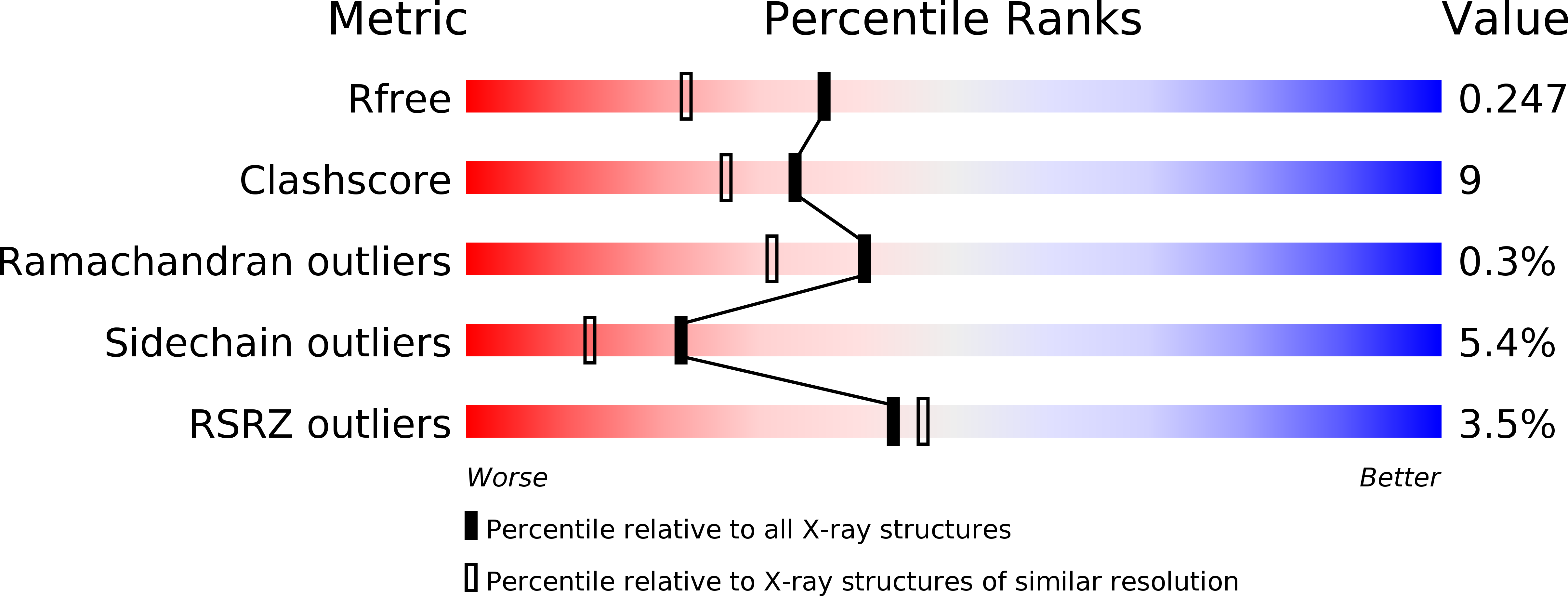
R-Value Free:
0.24
R-Value Work:
0.19
R-Value Observed:
0.19
Space Group:
P 1 21 1