Deposition Date
2006-02-20
Release Date
2006-08-15
Last Version Date
2024-10-30
Entry Detail
Biological Source:
Source Organism(s):
Pontellina plumata (Taxon ID: 239963)
Expression System(s):
Method Details:
Experimental Method:
Resolution:
2.10 Å
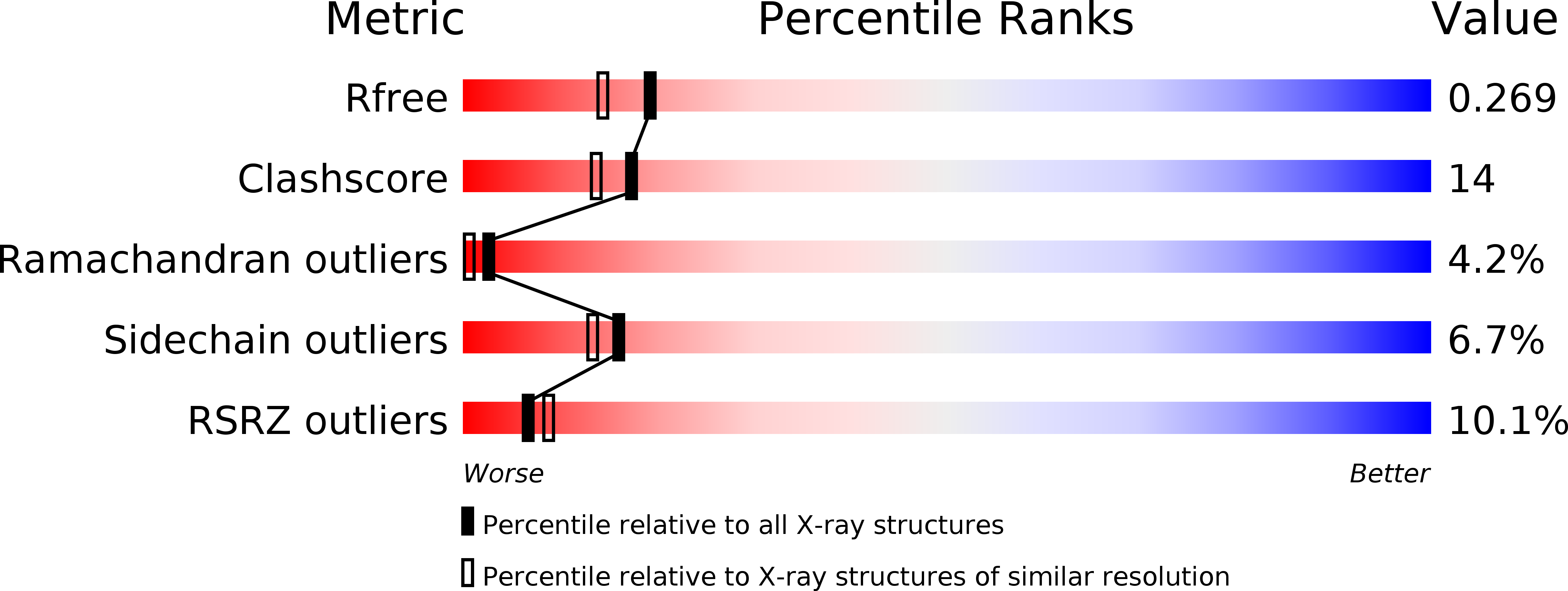
R-Value Free:
0.27
R-Value Work:
0.23
R-Value Observed:
0.23
Space Group:
P 32