2G2W image
Deposition Date 2006-02-16
Release Date 2006-07-04
Last Version Date 2024-11-20
Entry Detail
PDB ID:
2G2W
Title:
Crystal Structure of the SHV D104K Beta-lactamase/Beta-lactamase inhibitor protein (BLIP) complex
Biological Source:
Source Organism:
Method Details:
Experimental Method:
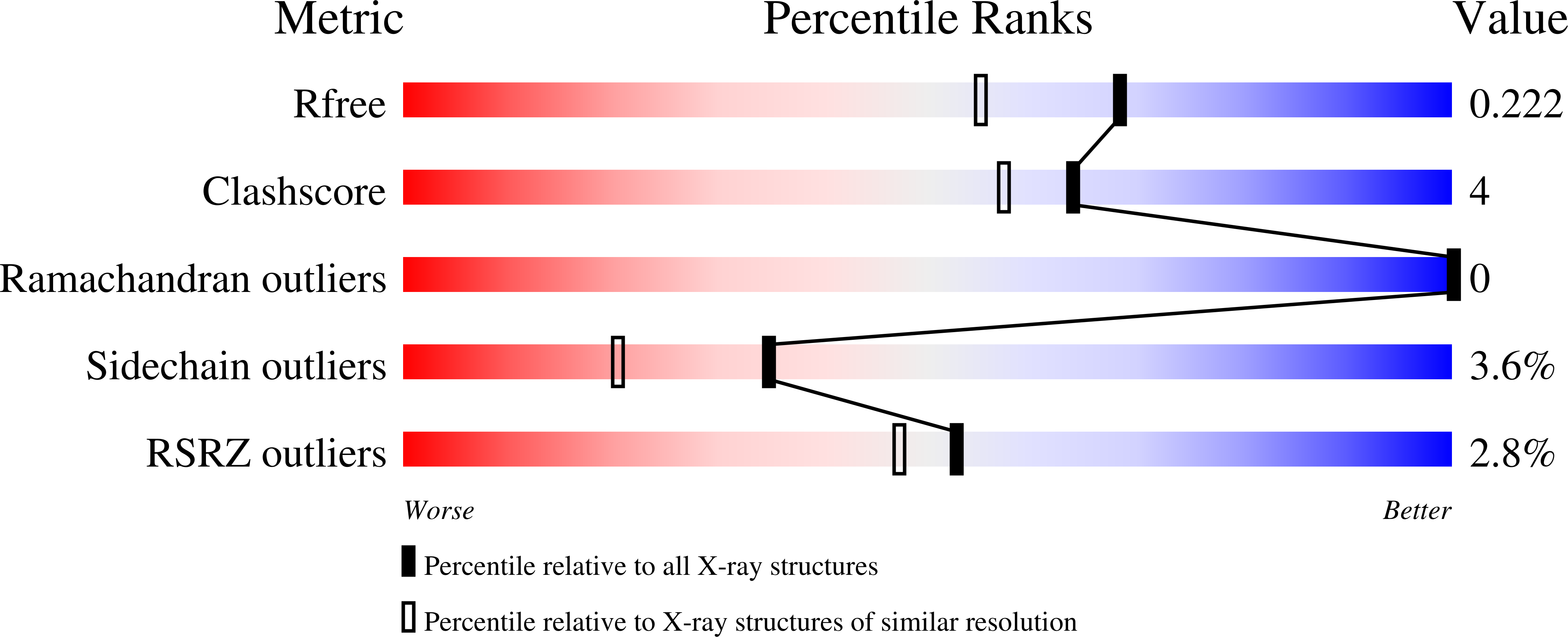
Resolution:
1.80 Å
R-Value Free:
0.22
R-Value Work:
0.17
R-Value Observed:
0.18
Space Group:
P 1
Macromolecular Entities
Polymer Type:polypeptide(L)
Molecule:Beta-lactamase SHV-1
Gene (Uniprot):bla
Mutations:D104K
Chain IDs:A
Chain Length:265
Number of Molecules:1
Biological Source:Klebsiella pneumoniae
Polymer Type:polypeptide(L)
Molecule:Beta-lactamase inhibitory protein
Chain IDs:B
Chain Length:165
Number of Molecules:1
Biological Source:Streptomyces clavuligerus
Primary Citation
Structural and Computational Characterization of the SHV-1 beta-Lactamase-beta-Lactamase Inhibitor Protein Interface.
J.Biol.Chem. 281 26745 26753 (2006)
PMID: 16809340 DOI: 10.1074/jbc.M603878200

Abstact

Beta-lactamase inhibitor protein (BLIP) binds a variety of class A beta-lactamases with affinities ranging from micromolar to picomolar. Whereas the TEM-1 and SHV-1 beta-lactamases are almost structurally identical, BLIP binds TEM-1 approximately 1000-fold tighter than SHV-1. Determining the underlying source of this affinity difference is important for understanding the molecular basis of beta-lactamase inhibition and mechanisms of protein-protein interface specificity and affinity. Here we present the 1.6A resolution crystal structure of SHV-1.BLIP. In addition, a point mutation was identified, SHV D104E, that increases SHV.BLIP binding affinity from micromolar to nanomolar. Comparison of the SHV-1.BLIP structure with the published TEM-1.BLIP structure suggests that the increased volume of Glu-104 stabilizes a key binding loop in the interface. Solution of the 1.8A SHV D104K.BLIP crystal structure identifies a novel conformation in which this binding loop is removed from the interface. Using these structural data, we evaluated the ability of EGAD, a program developed for computational protein design, to calculate changes in the stability of mutant beta-lactamase.BLIP complexes. Changes in binding affinity were calculated within an error of 1.6 kcal/mol of the experimental values for 112 mutations at the TEM-1.BLIP interface and within an error of 2.2 kcal/mol for 24 mutations at the SHV-1.BLIP interface. The reasonable success of EGAD in predicting changes in interface stability is a promising step toward understanding the stability of the beta-lactamase.BLIP complexes and computationally assisted design of tight binding BLIP variants.

Legend

Protein

Chemical

Disease

Primary Citation of related structures