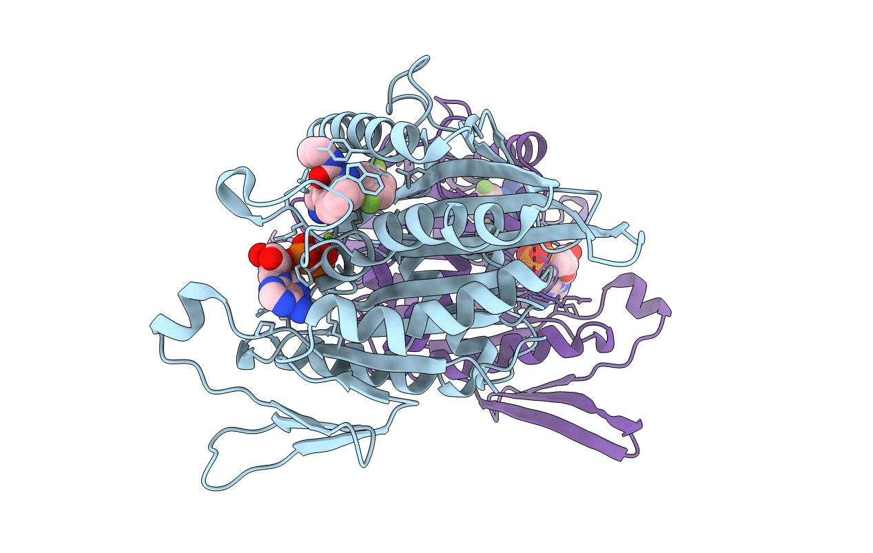
2G1Q image
Entry Detail
PDB ID:
2G1Q
Keywords:
Title:
crystal structure of KSP in complex with inhibitor 9h
Biological Source:
Source Organism:
Host Organism:
PDB Version:
Deposition Date:
2006-02-14
Release Date:
2006-10-03
Method Details:
Experimental Method:
Resolution:
2.51 Å
R-Value Free:
0.27
R-Value Work:
0.23
R-Value Observed:
0.23
Space Group:
P 21 21 21
Macromolecular Entities
Polymer Type:polypeptide(L)
Description:Kinesin-like protein KIF11
Chain IDs:A, B
Chain Length:368
Number of Molecules:2
Biological Source:Homo sapiens
Primary Citation
Kinesin spindle protein (KSP) inhibitors. Part 4: Structure-based design of 5-alkylamino-3,5-diaryl-4,5-dihydropyrazoles as potent, water-soluble inhibitors of the mitotic kinesin KSP.
Bioorg.Med.Chem.Lett. 16 3175 3179 (2006)
PMID: 16603356 DOI: 10.1016/j.bmcl.2006.03.040

Abstact

Molecular modeling in combination with X-ray crystallographic information was employed to identify a region of the kinesin spindle protein (KSP) binding site not fully utilized by our first generation inhibitors. We discovered that by appending a propylamine substituent at the C5 carbon of a dihydropyrazole core, we could effectively fill this unoccupied region of space and engage in a hydrogen-bonding interaction with the enzyme backbone. This change led to a second generation compound with increased potency, a 400-fold enhancement in aqueous solubility at pH 4, and improved dog pharmacokinetics relative to the first generation compound.

Legend

Protein

Chemical

Disease

Primary Citation of related structures