
Deposition Date
2006-02-13
Release Date
2006-05-23
Last Version Date
2024-05-29
Entry Detail
PDB ID:
2G0U
Keywords:
Title:
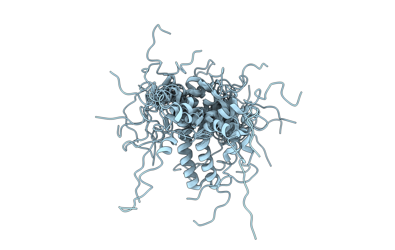
Solution Structure of Monomeric BsaL, the Type III Secretion Needle Protein of Burkholderia pseudomallei
Biological Source:
Source Organism(s):
Burkholderia pseudomallei (Taxon ID: 28450)
Expression System(s):
Method Details:
Experimental Method:
Conformers Calculated:
200
Conformers Submitted:
20
Selection Criteria:
structures with the least restraint violations,structures with the lowest energy
