2FZP image
Deposition Date 2006-02-10
Release Date 2006-03-28
Last Version Date 2024-02-14
Entry Detail
PDB ID:
2FZP
Keywords:
Title:
Crystal structure of the USP8 interaction domain of human NRDP1
Biological Source:
Source Organism:
Homo sapiens (Taxon ID: 9606)
Host Organism:
Method Details:
Experimental Method:
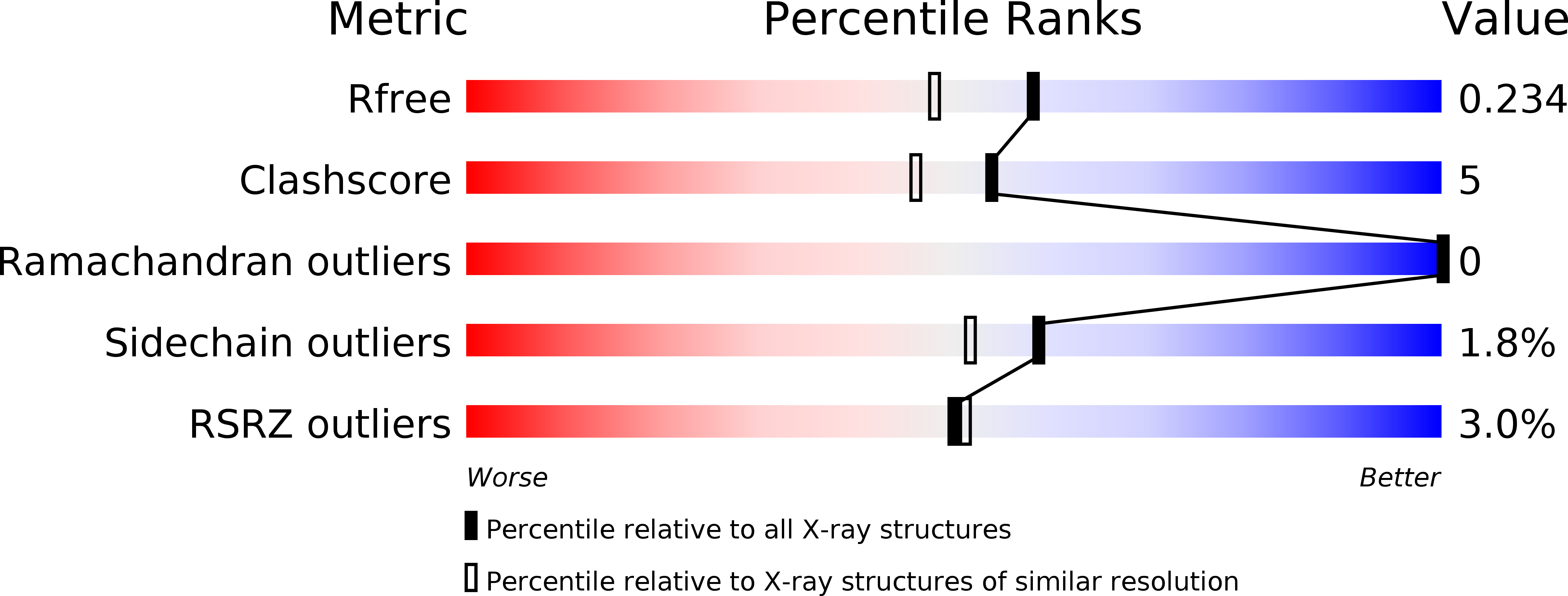
Resolution:
1.87 Å
R-Value Free:
0.23
R-Value Work:
0.16
R-Value Observed:
0.17
Space Group:
P 21 21 21
Macromolecular Entities
Polymer Type:polypeptide(L)
Molecule:ring finger protein 41 isoform 1
Gene (Uniprot):RNF41
Chain IDs:A
Chain Length:144
Number of Molecules:1
Biological Source:Homo sapiens
Primary Citation
Amino-terminal Dimerization, NRDP1-Rhodanese Interaction, and Inhibited Catalytic Domain Conformation of the Ubiquitin-specific Protease 8 (USP8).
J.Biol.Chem. 281 38061 38070 (2006)
PMID: 17035239 DOI: 10.1074/jbc.M606704200

Abstact

Ubiquitin-specific protease 8 (USP8) hydrolyzes mono and polyubiquitylated targets such as epidermal growth factor receptors and is involved in clathrin-mediated internalization. In 1182 residues, USP8 contains multiple domains, including coiled-coil, rhodanese, and catalytic domains. We report the first high-resolution crystal structures of these domains and discuss their implications for USP8 function. The amino-terminal domain is a homodimer with a novel fold. It is composed of two five-helix bundles, where the first helices are swapped, and carboxyl-terminal helices are extended in an antiparallel fashion. The structure of the rhodanese domain, determined in complex with the E3 ligase NRDP1, reveals the canonical rhodanese fold but with a distorted primordial active site. The USP8 recognition domain of NRDP1 has a novel protein fold that interacts with a conserved peptide loop of the rhodanese domain. A consensus sequence of this loop is found in other NRDP1 targets, suggesting a common mode of interaction. The structure of the carboxyl-terminal catalytic domain of USP8 exhibits the conserved tripartite architecture but shows unique traits. Notably, the active site, including the ubiquitin binding pocket, is in a closed conformation, incompatible with substrate binding. The presence of a zinc ribbon subdomain near the ubiquitin binding site further suggests a polyubiquitin-specific binding site and a mechanism for substrate induced conformational changes.

Legend

Protein

Chemical

Disease

Primary Citation of related structures