Deposition Date
2006-02-09
Release Date
2006-10-03
Last Version Date
2023-08-30
Entry Detail
PDB ID:
2FZD
Keywords:
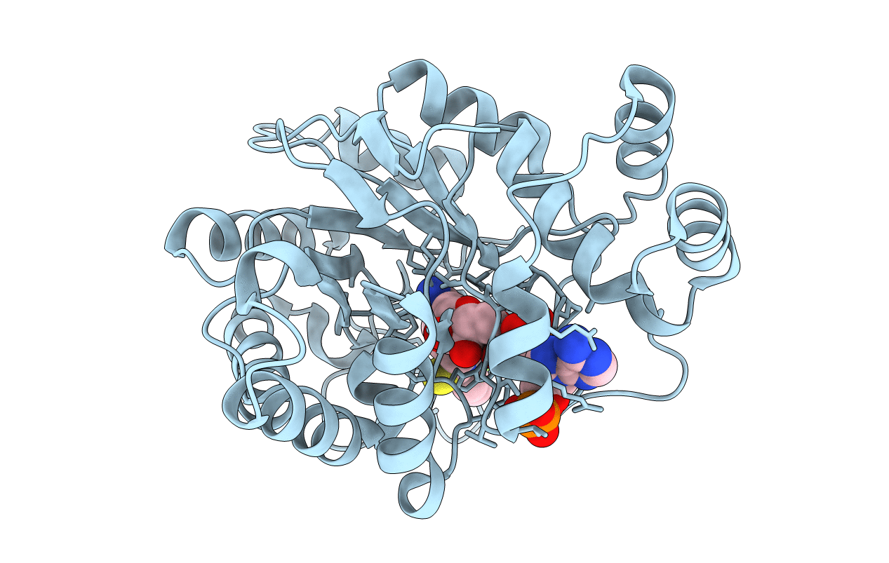
Title:
Human aldose reductase complexed with tolrestat at 1.08 A resolution.
Biological Source:
Source Organism(s):
Homo sapiens (Taxon ID: 9606)
Expression System(s):
Method Details:
Experimental Method:
Resolution:
1.08 Å
R-Value Free:
0.13
R-Value Work:
0.11
R-Value Observed:
0.11
Space Group:
P 1 21 1