
Deposition Date
2006-02-06
Release Date
2006-03-07
Last Version Date
2023-08-30
Entry Detail
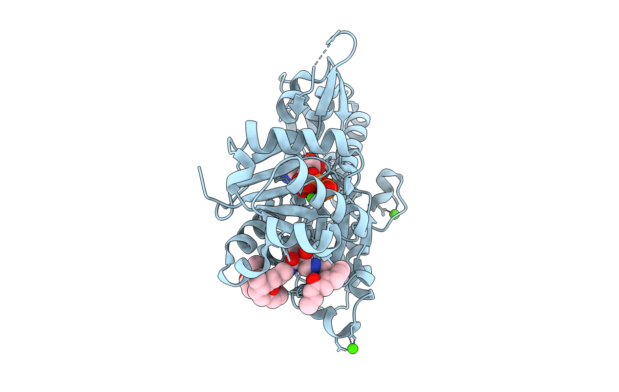
PDB ID:
2FXU
Keywords:
Title:
X-ray Structure of Bistramide A- Actin Complex at 1.35 A resolution.
Biological Source:
Source Organism(s):
Oryctolagus cuniculus (Taxon ID: 9986)
Method Details:
Experimental Method:
Resolution:
1.35 Å
R-Value Free:
0.20
R-Value Work:
0.17
R-Value Observed:
0.17
Space Group:
C 1 2 1
