2FXT image
Deposition Date 2006-02-06
Release Date 2007-02-06
Last Version Date 2024-02-14
Entry Detail
PDB ID:
2FXT
Title:
Crystal Structure of Yeast Tim44
Biological Source:
Source Organism:
Host Organism:
Method Details:
Experimental Method:
Resolution:
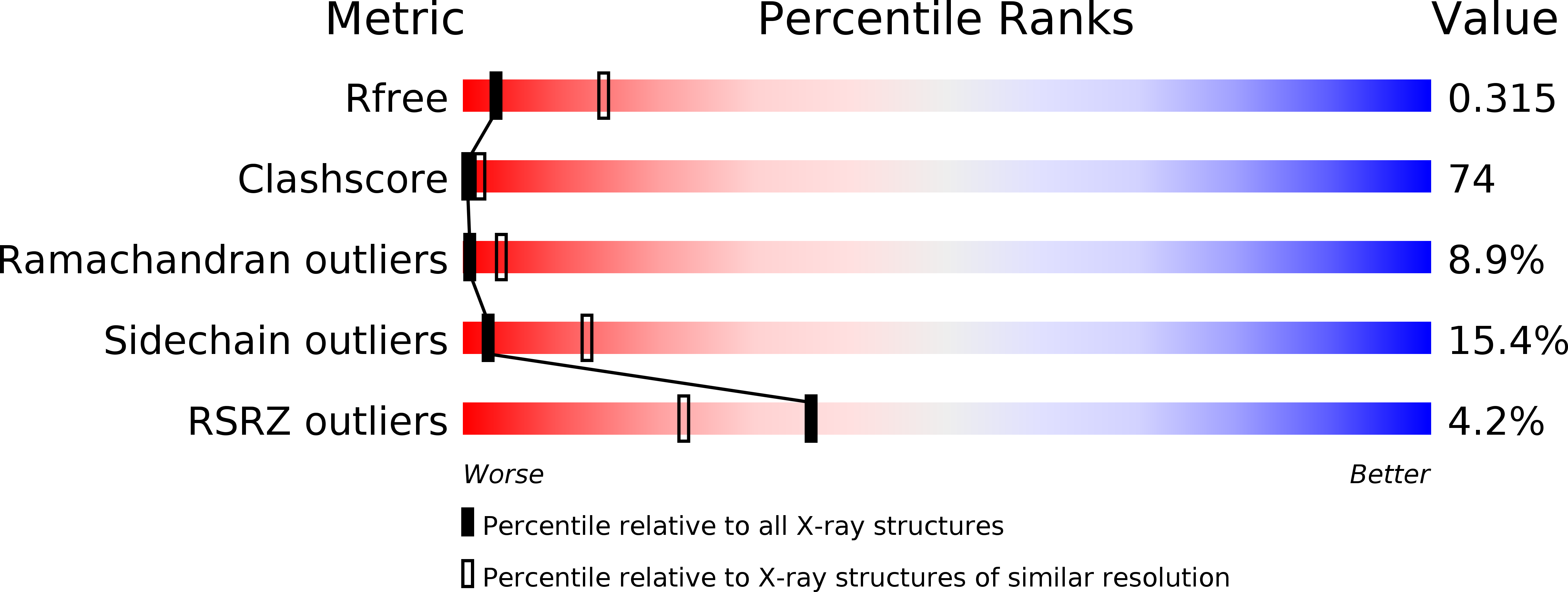
3.20 Å
R-Value Free:
0.36
R-Value Work:
0.31
R-Value Observed:
0.31
Space Group:
P 63 2 2
Macromolecular Entities
Structures with similar UniProt ID
Protein Blast
Polymer Type:polypeptide(L)
Molecule:Import inner membrane translocase subunit TIM44
Gene (Uniprot):TIM44
Chain IDs:A
Chain Length:192
Number of Molecules:1
Biological Source:Saccharomyces cerevisiae
Ligand Molecules
Primary Citation
Crystal Structure of Yeast Mitochondrial Peripheral Membrane Protein Tim44p C-terminal Domain.
J.Mol.Biol. 359 798 804 (2006)
PMID: 16647716 DOI: 10.1016/j.jmb.2006.04.020

Abstact

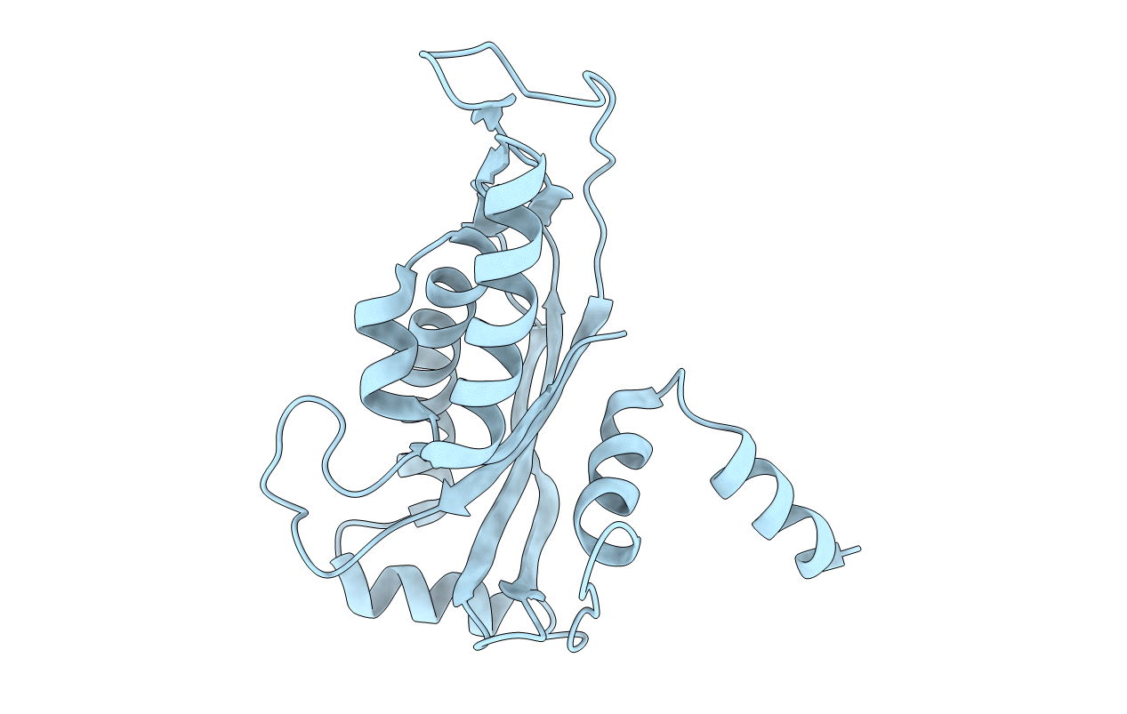
The protein transports from the cell cytosol to the mitochondria matrix are carried out by the translocase of the outer membrane (TOM) complex and the translocase of the inner membrane (TIM) complexes. Tim44p is an essential mitochondrial peripheral membrane protein and a major component of TIM23 translocon. Tim44p can tightly associate with the inner mitochondrial membrane. To investigate the mechanism by which Tim44p functions in the TIM23 translocon to deliver the mitochondrial protein precursors, we have determined the crystal structure of the yeast Tim44p C-terminal domain to 3.2A resolution using the MAD method. The Tim44p C-terminal domain forms a monomer in the crystal structure and contains six alpha-helices and four antiparallel beta-strands. A large hydrophobic pocket was identified on the Tim44p structure surface. The N-terminal helix A1 is positively charged and the helix A1 protrudes out from the Tim44p main body.

Legend

Protein

Chemical

Disease

Primary Citation of related structures
Feedback Form
Name
Email
Institute
Feedback