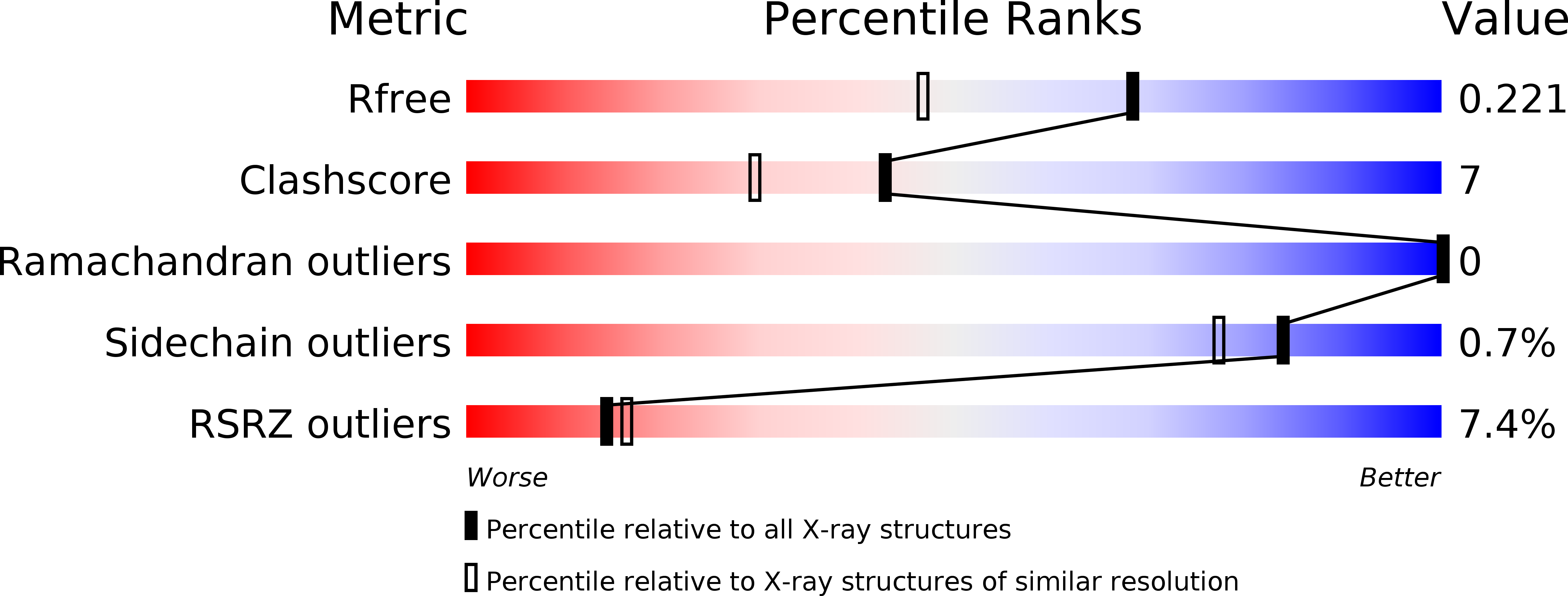
2FWV image
Deposition Date 2006-02-03
Release Date 2006-08-03
Last Version Date 2024-03-13
Entry Detail
PDB ID:
2FWV
Title:
Crystal Structure of Rv0813
Biological Source:
Source Organism:
Host Organism:
Method Details:
Experimental Method:
Resolution:
1.70 Å
R-Value Free:
0.22
R-Value Work:
0.18
R-Value Observed:
0.18
Space Group:
I 41 2 2
Macromolecular Entities
Polymer Type:polypeptide(L)
Molecule:hypothetical protein MtubF_01000852
Chain IDs:A
Chain Length:226
Number of Molecules:1
Biological Source:Mycobacterium tuberculosis
Primary Citation
The Crystal Structure of Rv0813c from Mycobacterium tuberculosis Reveals a New Family of Fatty Acid-Binding Protein-Like Proteins in Bacteria
J.Bacteriol. 189 1899 1904 (2007)
PMID: 17172346 DOI: 10.1128/JB.01435-06

Abstact

The gene Rv0813c from Mycobacterium tuberculosis, which codes for a hypothetical protein of unknown function, is conserved within the order Actinomycetales but absent elsewhere. The crystal structure of Rv0813c reveals a new family of proteins that resemble the fatty acid-binding proteins (FABPs) found in eukaryotes. Rv0813c adopts the 10-stranded beta-barrel fold typical of FABPs but lacks the double-helix insert that covers the entry to the binding site in the eukaryotic proteins. The barrel encloses a deep cavity, at the bottom of which a small cyclic ligand was found to bind to the hydroxyl group of Tyr192. This residue is part of a conserved Arg-X-Tyr motif much like the triad that binds the carboxylate group of fatty acids in FABPs. Most of the residues forming the internal surface of the cavity are conserved in homologous protein sequences found in CG-rich prokaryotes, strongly suggesting that Rv0813c is a member of a new family of bacterial FABP-like proteins that may have roles in the recognition, transport, and/or storage of small molecules in the bacterial cytosol.

Legend

Protein

Chemical

Disease

Primary Citation of related structures