Deposition Date
2006-01-31
Release Date
2007-02-06
Last Version Date
2024-04-03
Entry Detail
PDB ID:
2FVY
Keywords:
Title:
High Resolution Glucose Bound Crystal Structure of GGBP
Biological Source:
Source Organism(s):
Escherichia coli (Taxon ID: 562)
Expression System(s):
Method Details:
Experimental Method:
Resolution:
0.92 Å
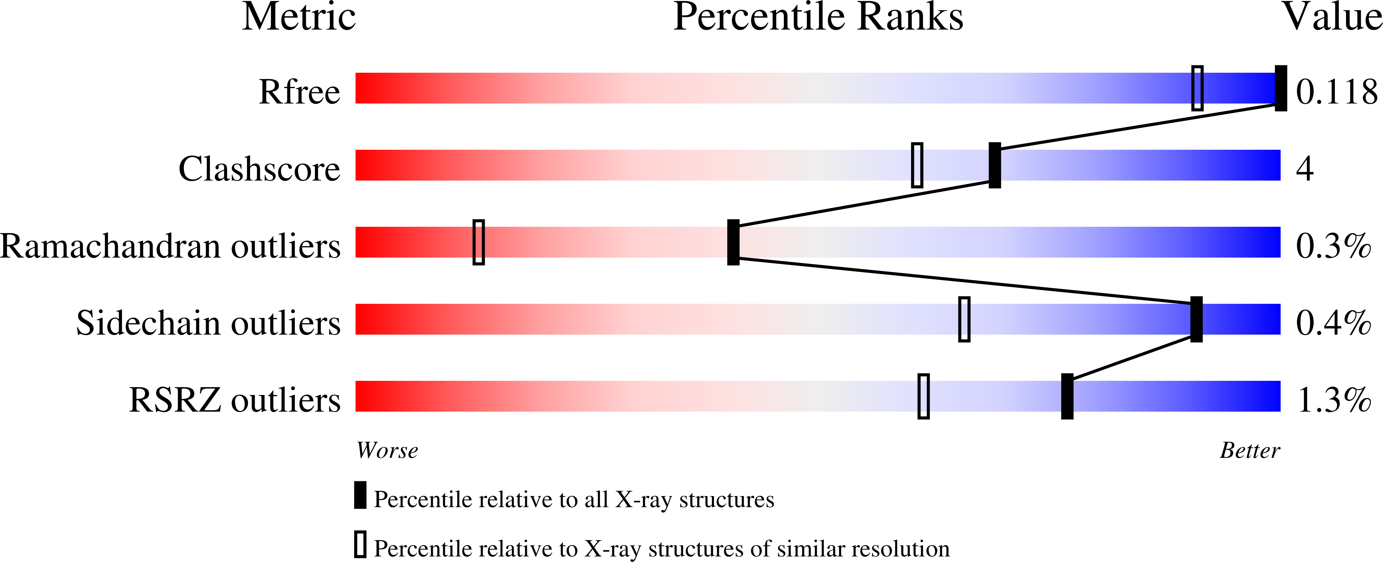
R-Value Free:
0.12
R-Value Work:
0.11
R-Value Observed:
0.11
Space Group:
C 1 2 1