2FVM image
Deposition Date 2006-01-31
Release Date 2006-03-14
Last Version Date 2023-11-15
Entry Detail
PDB ID:
2FVM
Keywords:
Title:
Crystal structure of dihydropyrimidinase from Saccharomyces kluyveri in complex with the reaction product N-carbamyl-beta-alanine
Biological Source:
Source Organism:
Host Organism:
Method Details:
Experimental Method:
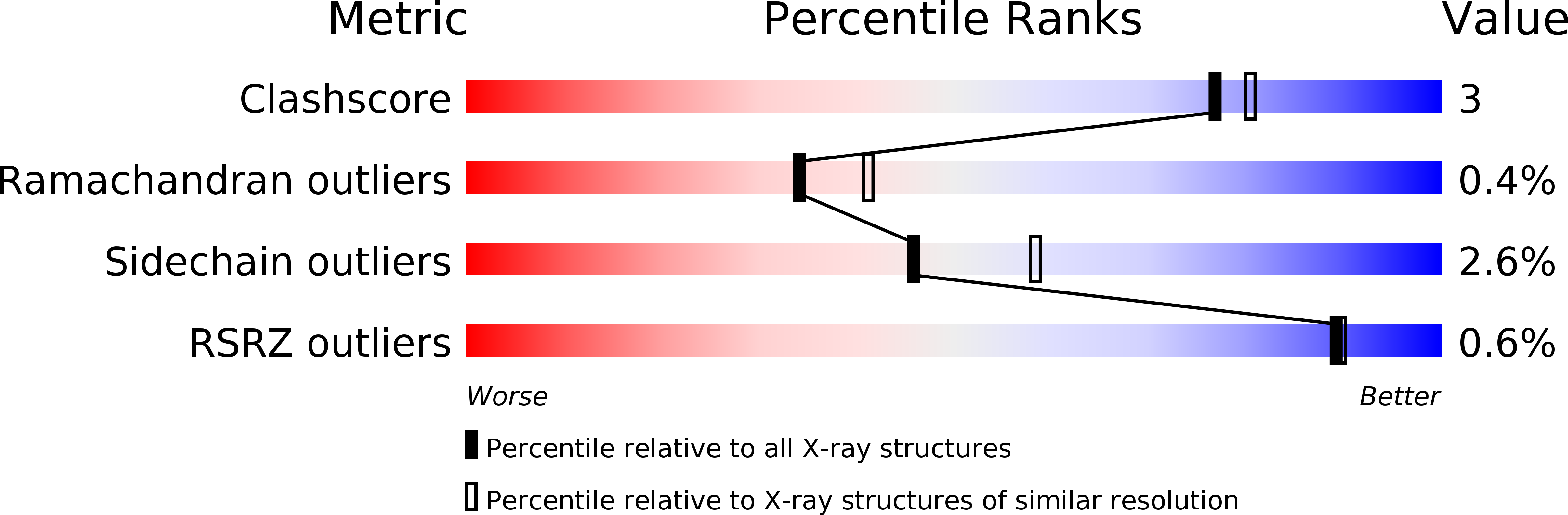
Resolution:
2.45 Å
R-Value Free:
0.23
R-Value Work:
0.18
R-Value Observed:
0.18
Space Group:
P 1 21 1
Macromolecular Entities
Polymer Type:polypeptide(L)
Molecule:dihydropyrimidinase
Gene (Uniprot):PYD2
Chain IDs:A, B, C, D
Chain Length:559
Number of Molecules:4
Biological Source:Lachancea kluyveri
Modified Residue
Compound ID Chain ID Parent Comp ID Details 2D Image
KCX A LYS LYSINE NZ-CARBOXYLIC ACID
Primary Citation
The Crystal Structures of Dihydropyrimidinases Reaffirm the Close Relationship between Cyclic Amidohydrolases and Explain Their Substrate Specificity.
J.Biol.Chem. 281 13762 13776 (2006)
PMID: 16517602 DOI: 10.1074/jbc.M513266200

Abstact

In eukaryotes, dihydropyrimidinase catalyzes the second step of the reductive pyrimidine degradation, the reversible hydrolytic ring opening of dihydropyrimidines. Here we describe the three-dimensional structures of dihydropyrimidinase from two eukaryotes, the yeast Saccharomyces kluyveri and the slime mold Dictyostelium discoideum, determined and refined to 2.4 and 2.05 angstroms, respectively. Both enzymes have a (beta/alpha)8-barrel structural core embedding the catalytic di-zinc center, which is accompanied by a smaller beta-sandwich domain. Despite loop-forming insertions in the sequence of the yeast enzyme, the overall structures and architectures of the active sites of the dihydropyrimidinases are strikingly similar to each other, as well as to those of hydantoinases, dihydroorotases, and other members of the amidohydrolase superfamily of enzymes. However, formation of the physiologically relevant tetramer shows subtle but nonetheless significant differences. The extension of one of the sheets of the beta-sandwich domain across a subunit-subunit interface in yeast dihydropyrimidinase underlines its closer evolutionary relationship to hydantoinases, whereas the slime mold enzyme shows higher similarity to the noncatalytic collapsin-response mediator proteins involved in neuron development. Catalysis is expected to follow a dihydroorotase-like mechanism but in the opposite direction and with a different substrate. Complexes with dihydrouracil and N-carbamyl-beta-alanine obtained for the yeast dihydropyrimidinase reveal the mode of substrate and product binding and allow conclusions about what determines substrate specificity, stereoselectivity, and the reaction direction among cyclic amidohydrolases.

Legend

Protein

Chemical

Disease

Primary Citation of related structures