2FUF image
Deposition Date 2006-01-26
Release Date 2006-08-22
Last Version Date 2024-10-09
Entry Detail
PDB ID:
2FUF
Title:
Crystal structure of the SV40 large T antigen origin-binding domain
Biological Source:
Source Organism(s):
Expression System(s):
Method Details:
Experimental Method:
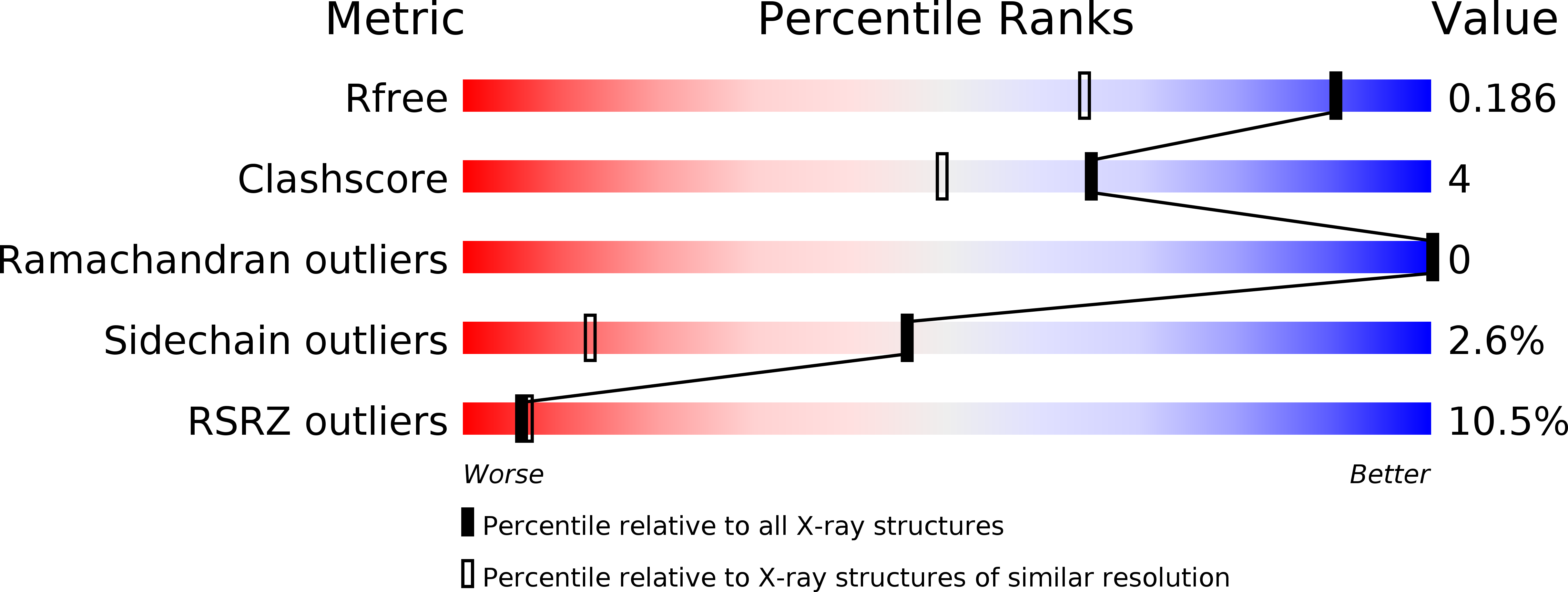
Resolution:
1.45 Å
R-Value Free:
0.18
R-Value Work:
0.16
R-Value Observed:
0.17
Space Group:
P 65
Macromolecular Entities
Structures with similar UniProt ID
Protein Blast
Polymer Type:polypeptide(L)
Molecule:Large T antigen
Chain IDs:A
Chain Length:134
Number of Molecules:1
Biological Source:Simian virus 40
Modified Residue
Compound ID Chain ID Parent Comp ID Details 2D Image
MSE A MET SELENOMETHIONINE
Ligand Molecules
Primary Citation
Crystal structure of the simian virus 40 large T-antigen origin-binding domain.
J.Virol. 80 4304 4312 (2006)
PMID: 16611889 DOI: 10.1128/JVI.80.9.4304-4312.2006

Abstact

The origins of replication of DNA tumor viruses have a highly conserved feature, namely, multiple binding sites for their respective initiator proteins arranged as inverted repeats. In the 1.45-angstroms crystal structure of the simian virus 40 large T-antigen (T-ag) origin-binding domain (obd) reported herein, T-ag obd monomers form a left-handed spiral with an inner channel of 30 angstroms having six monomers per turn. The inner surface of the spiral is positively charged and includes residues known to bind DNA. Residues implicated in hexamerization of full-length T-ag are located at the interface between adjacent T-ag obd monomers. These data provide a high-resolution model of the hexamer of origin-binding domains observed in electron microscopy studies and allow the obd's to be oriented relative to the hexamer of T-ag helicase domains to which they are connected.

Legend

Protein

Chemical

Disease

Primary Citation of related structures
Feedback Form
Name
Email
Institute
Feedback