2FUE image
Deposition Date 2006-01-26
Release Date 2006-03-21
Last Version Date 2024-10-30
Entry Detail
PDB ID:
2FUE
Keywords:
Title:
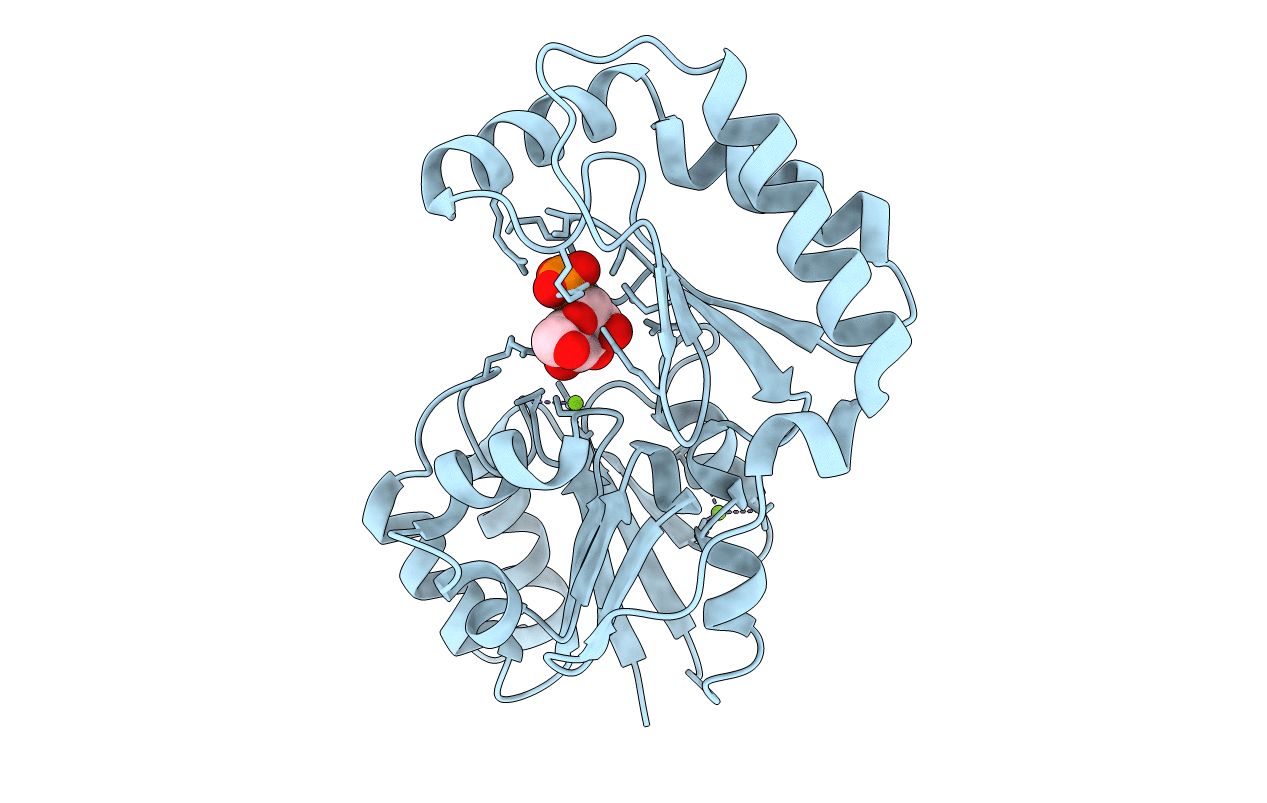
Human alpha-Phosphomannomutase 1 with D-mannose 1-phosphate and Mg2+ cofactor bound
Biological Source:
Source Organism:
Homo sapiens (Taxon ID: 9606)
Host Organism:
Method Details:
Experimental Method:
Resolution:
1.75 Å
R-Value Free:
0.24
R-Value Work:
0.20
R-Value Observed:
0.20
Space Group:
P 43 21 2
Macromolecular Entities
Polymer Type:polypeptide(L)
Molecule:Phosphomannomutase 1
Gene (Uniprot):PMM1
Chain IDs:A
Chain Length:262
Number of Molecules:1
Biological Source:Homo sapiens
Modified Residue
Compound ID Chain ID Parent Comp ID Details 2D Image
MSE A MET SELENOMETHIONINE
Primary Citation
The X-ray crystal structures of human alpha-phosphomannomutase 1 reveal the structural basis of congenital disorder of glycosylation type 1a.
J.Biol.Chem. 281 14918 14926 (2006)
PMID: 16540464 DOI: 10.1074/jbc.M601505200

Abstact

Congenital disorder of glycosylation type 1a (CDG-1a) is a congenital disease characterized by severe defects in nervous system development. It is caused by mutations in alpha-phosphomannomutase (of which there are two isozymes, alpha-PMM1 and alpha-PPM2). Here we report the x-ray crystal structures of human alpha-PMM1 in the open conformation, with and without the bound substrate, alpha-D-mannose 1-phosphate. Alpha-PMM1, like most haloalkanoic acid dehalogenase superfamily (HADSF) members, consists of two domains, the cap and core, which open to bind substrate and then close to provide a solvent-exclusive environment for catalysis. The substrate phosphate group is observed at a positively charged site of the cap domain, rather than at the core domain phosphoryl-transfer site defined by the Asp(19) nucleophile and Mg(2+) cofactor. This suggests that substrate binds first to the cap and then is swept into the active site upon cap closure. The orientation of the acid/base residue Asp(21) suggests that alpha-phosphomannomutase (alpha-PMM) uses a different method of protecting the aspartylphosphate from hydrolysis than the HADSF member beta-phosphoglucomutase. It is hypothesized that the electrostatic repulsion of positive charges at the interface of the cap and core domains stabilizes alpha-PMM1 in the open conformation and that the negatively charged substrate binds to the cap, thereby facilitating its closure over the core domain. The two isozymes, alpha-PMM1 and alpha-PMM2, are shown to have a conserved active-site structure and to display similar kinetic properties. Analysis of the known mutation sites in the context of the structures reveals the genotype-phenotype relationship underlying CDG-1a.

Legend

Protein

Chemical

Disease

Primary Citation of related structures