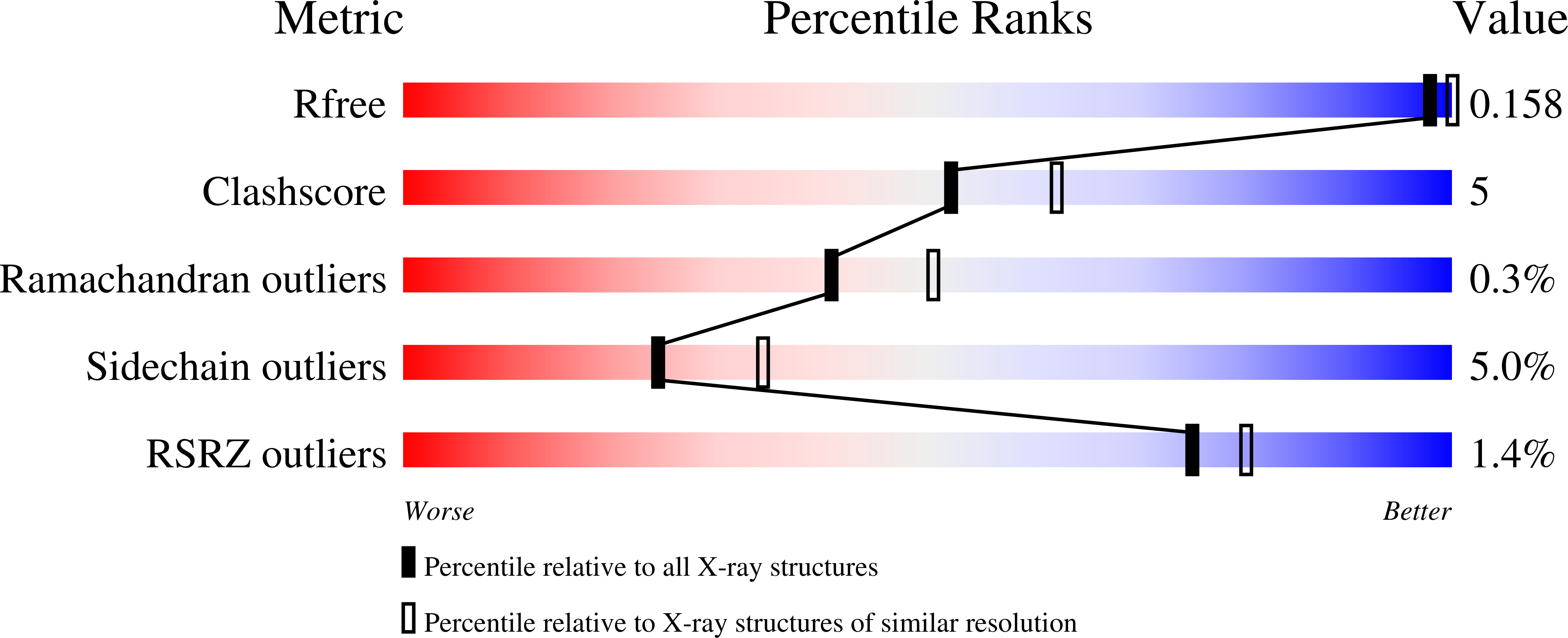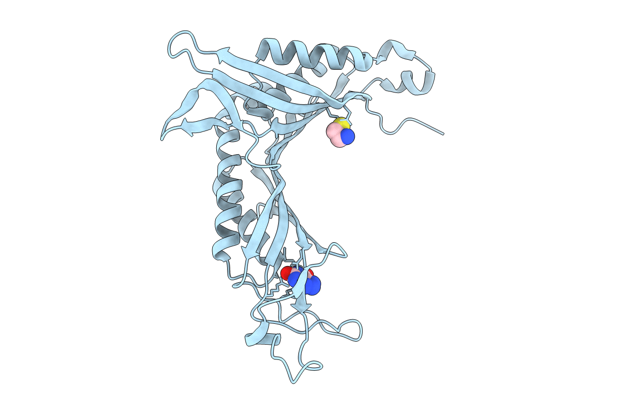
2FUB image
Deposition Date 2006-01-26
Release Date 2006-02-14
Last Version Date 2024-10-30
Entry Detail
PDB ID:
2FUB
Keywords:
Title:
Crystal structure of urate oxidase at 140 MPa
Biological Source:
Source Organism:
Method Details:
Experimental Method:
Resolution:
2.30 Å
R-Value Free:
0.21
R-Value Work:
0.16
R-Value Observed:
0.16
Space Group:
I 2 2 2
Macromolecular Entities
Polymer Type:polypeptide(L)
Molecule:Uricase
Gene (Uniprot):uaZ
Chain IDs:A
Chain Length:301
Number of Molecules:1
Biological Source:Aspergillus flavus
Primary Citation
High pressure macromolecular crystallography: The 140-MPa crystal structure at 2.3 A resolution of urate oxidase, a 135-kDa tetrameric assembly
Biochim.Biophys.Acta 1764 391 397 (2006)
PMID: 16478683 DOI: 10.1016/j.bbapap.2006.01.006

Abstact

We report the three-dimensional structure determined by high-pressure macromolecular crystallography (HPMX) of a 135-kDa homo-tetrameric enzyme, urate oxidase from Aspergillus flavus complexed with its potent inhibitor 8-azaxanthin. Urate oxidase crystals are quite sensitive to pressure, as three-dimensional order is lost at about 180 MPa. A highly complete 2.3 A resolution data set was collected at 140 MPa, close to the critical pressure. Crystal structures at atmospheric pressure and at high pressure were refined in the orthorhombic space group I222 with final crystallographic R factors 14.1% and 16.1%, respectively. The effect of pressure on temperature factors, ordered water molecules, hydrogen bond lengths, contacts, buried surface areas as well as cavity volume was investigated. Results suggest that the onset of disruption of the tetrameric assembly by pressure has been captured in the crystalline state.

Legend

Protein

Chemical

Disease

Primary Citation of related structures