Deposition Date
2006-01-26
Release Date
2006-05-16
Last Version Date
2024-03-13
Entry Detail
PDB ID:
2FU4
Keywords:
Title:
Crystal Structure of the DNA binding domain of E.coli FUR (Ferric Uptake Regulator)
Biological Source:
Source Organism(s):
Escherichia coli (Taxon ID: 511693)
Expression System(s):
Method Details:
Experimental Method:
Resolution:
1.80 Å
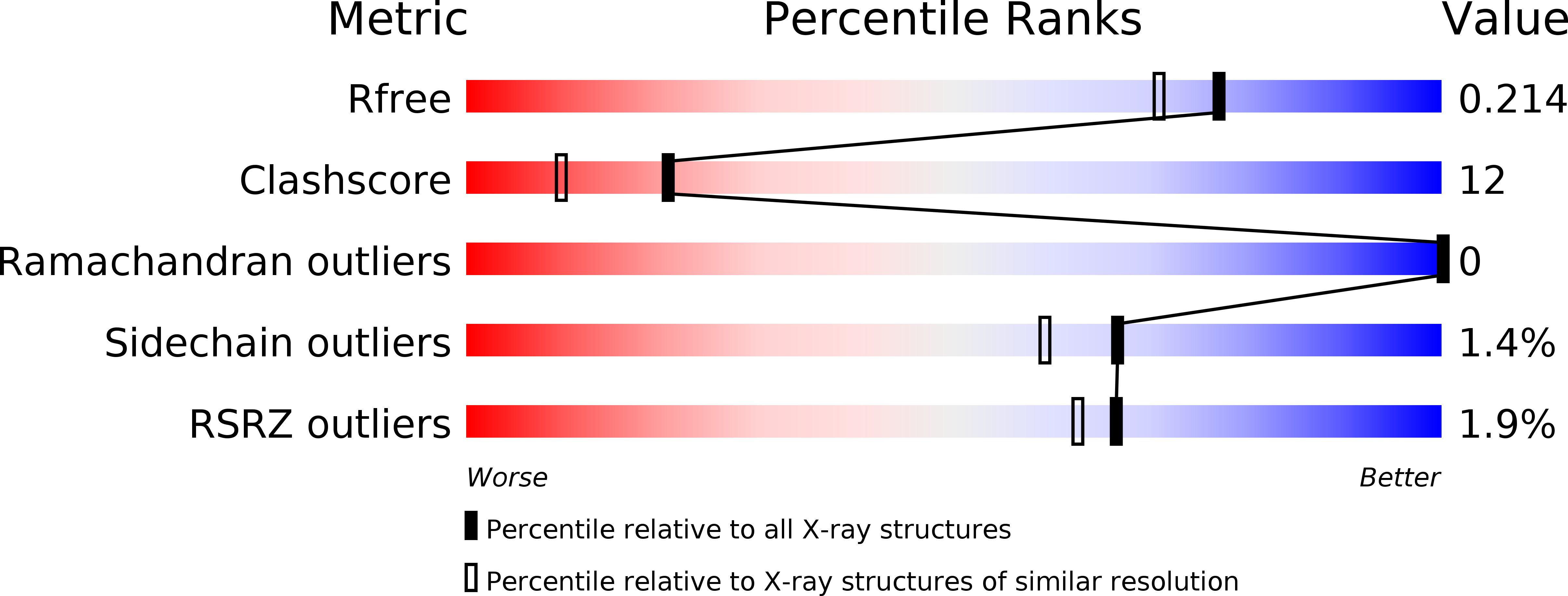
R-Value Free:
0.21
R-Value Work:
0.17
R-Value Observed:
0.17
Space Group:
P 21 21 2