2FTS image
Deposition Date 2006-01-24
Release Date 2006-03-14
Last Version Date 2024-02-14
Entry Detail
PDB ID:
2FTS
Title:
Crystal structure of the glycine receptor-gephyrin complex
Biological Source:
Source Organism:
Method Details:
Experimental Method:
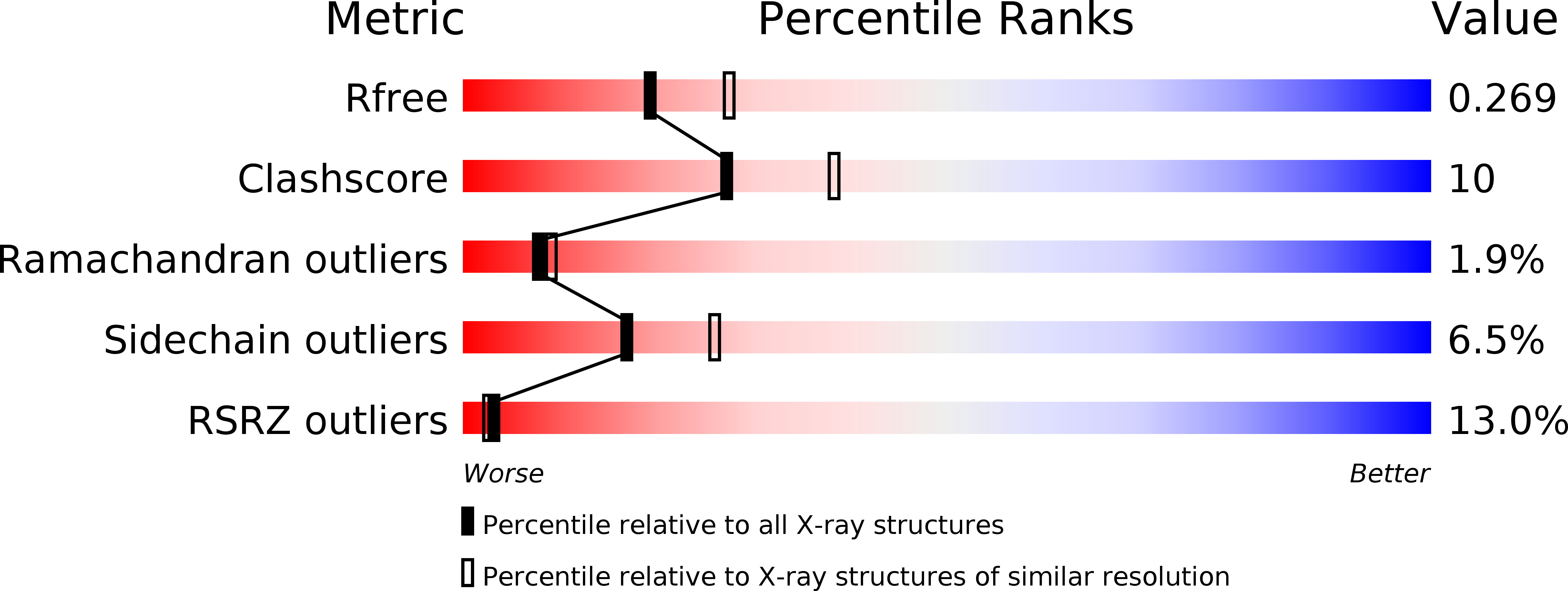
Resolution:
2.41 Å
R-Value Free:
0.27
R-Value Work:
0.19
Space Group:
C 2 2 21
Macromolecular Entities
Polymer Type:polypeptide(L)
Molecule:gephyrin
Gene (Uniprot):Gphn
Chain IDs:A
Chain Length:419
Number of Molecules:1
Biological Source:Rattus norvegicus
Polymer Type:polypeptide(L)
Molecule:Glycine receptor beta chain precursor
Gene (Uniprot):Glrb
Chain IDs:B (auth: P)
Chain Length:13
Number of Molecules:1
Biological Source:Rattus norvegicus
Primary Citation
Deciphering the structural framework of glycine receptor anchoring by gephyrin.
Embo J. 25 1385 1395 (2006)
PMID: 16511563 DOI: 10.1038/sj.emboj.7601029

Abstact

Glycine is the major inhibitory neurotransmitter in the spinal cord and brain stem. Gephyrin is required to achieve a high concentration of glycine receptors (GlyRs) in the postsynaptic membrane, which is crucial for efficient glycinergic signal transduction. The interaction between gephyrin and the GlyR involves the E-domain of gephyrin and a cytoplasmic loop located between transmembrane segments three and four of the GlyR beta subunit. Here, we present crystal structures of the gephyrin E-domain with and without the GlyR beta-loop at 2.4 and 2.7 A resolutions, respectively. The GlyR beta-loop is bound in a symmetric 'key and lock' fashion to each E-domain monomer in a pocket adjacent to the dimer interface. Structure-guided mutagenesis followed by in vitro binding and in vivo colocalization assays demonstrate that a hydrophobic interaction formed by Phe 330 of gephyrin and Phe 398 and Ile 400 of the GlyR beta-loop is crucial for binding.

Legend

Protein

Chemical

Disease

Primary Citation of related structures