2FS2 image
Deposition Date 2006-01-20
Release Date 2006-02-07
Last Version Date 2024-10-16
Entry Detail
PDB ID:
2FS2
Keywords:
Title:
Structure of the E. coli PaaI protein from the phyenylacetic acid degradation operon
Biological Source:
Source Organism:
Escherichia coli (Taxon ID: 562)
Host Organism:
Method Details:
Experimental Method:
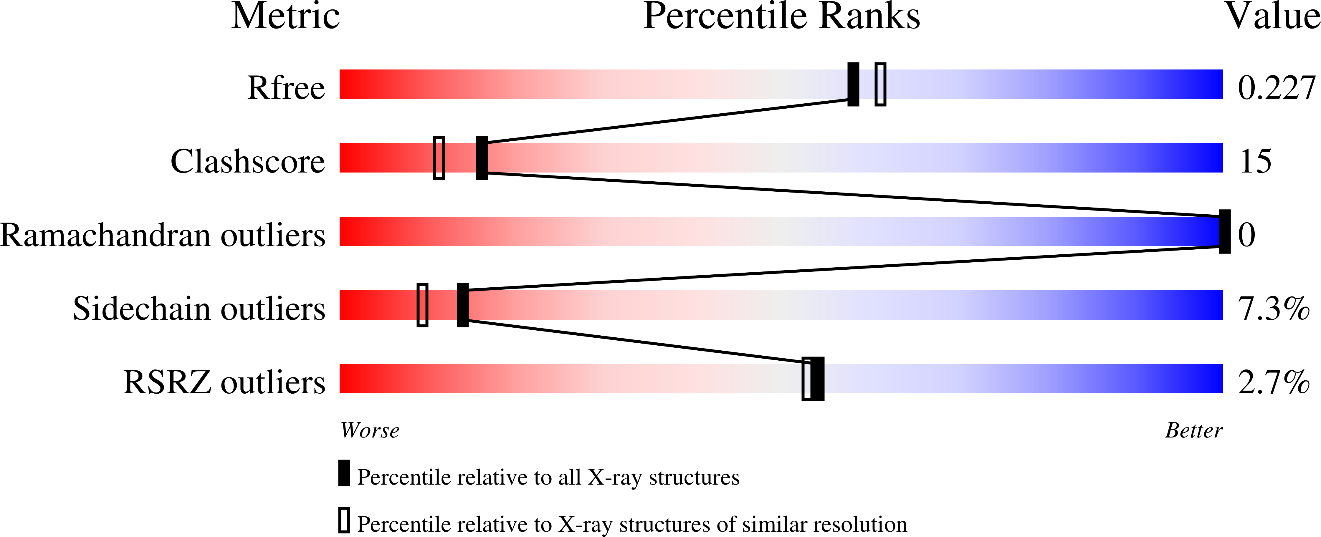
Resolution:
2.00 Å
R-Value Free:
0.23
R-Value Work:
0.18
Space Group:
P 31 2 1
Macromolecular Entities
Polymer Type:polypeptide(L)
Molecule:Phenylacetic acid degradation protein paaI
Gene (Uniprot):paaI
Chain IDs:A, B
Chain Length:151
Number of Molecules:2
Biological Source:Escherichia coli
Modified Residue
Compound ID Chain ID Parent Comp ID Details 2D Image
MSE A MET SELENOMETHIONINE
Ligand Molecules
Primary Citation
Structure, Function, and Mechanism of the Phenylacetate Pathway Hot Dog-fold Thioesterase PaaI
J.Biol.Chem. 281 11028 11038 (2006)
PMID: 16464851 DOI: 10.1074/jbc.M513896200

Abstact

The structure and biochemical function of the hot dog-fold thioesterase PaaI operative in the aerobic phenylacetate degradation pathway are examined. PaaI showed modest activity with phenylacetyl-coenzyme A, suggestive of a role in coenzyme A release from this pathway intermediate in the event of limiting downstream pathway enzymes. Minimal activity was observed with aliphatic acyl-coenzyme A thioesters, which ruled out PaaI function in the lower phenylacetate pathway. PaaI was most active with ring-hydroxylated phenylacetyl-coenzyme A thioesters. The x-ray crystal structure of the Escherichia coli thioesterase is reported and analyzed to define the structural basis of substrate recognition and catalysis. The contributions of catalytic and substrate binding residues, thus, identified were examined through steady-state kinetic analysis of site-directed mutant proteins.

Legend

Protein

Chemical

Disease

Primary Citation of related structures