Deposition Date
2006-01-18
Release Date
2006-04-04
Last Version Date
2024-04-03
Entry Detail
PDB ID:
2FR0
Keywords:
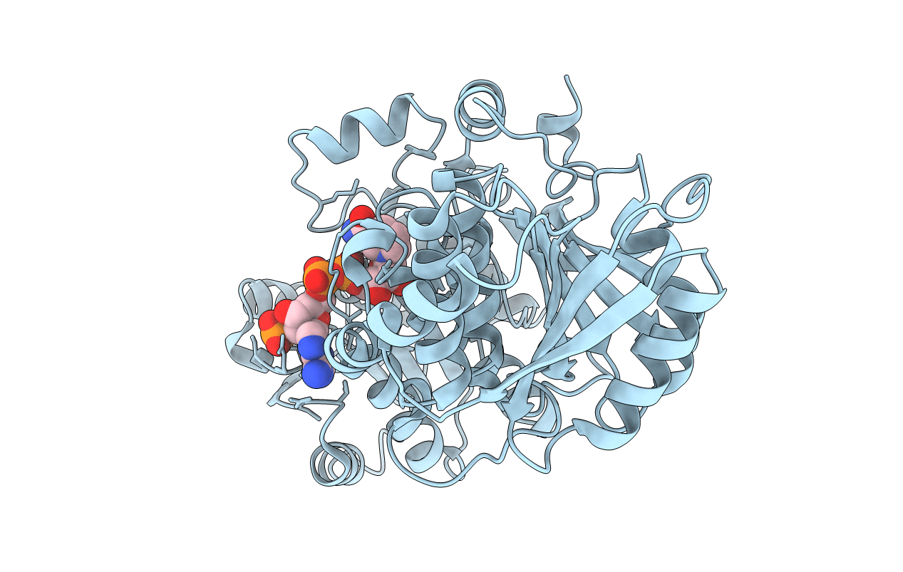
Title:
The first ketoreductase of the erythromycin synthase (crystal form 1)
Biological Source:
Source Organism(s):
Saccharopolyspora erythraea (Taxon ID: 1836)
Expression System(s):
Method Details:
Experimental Method:
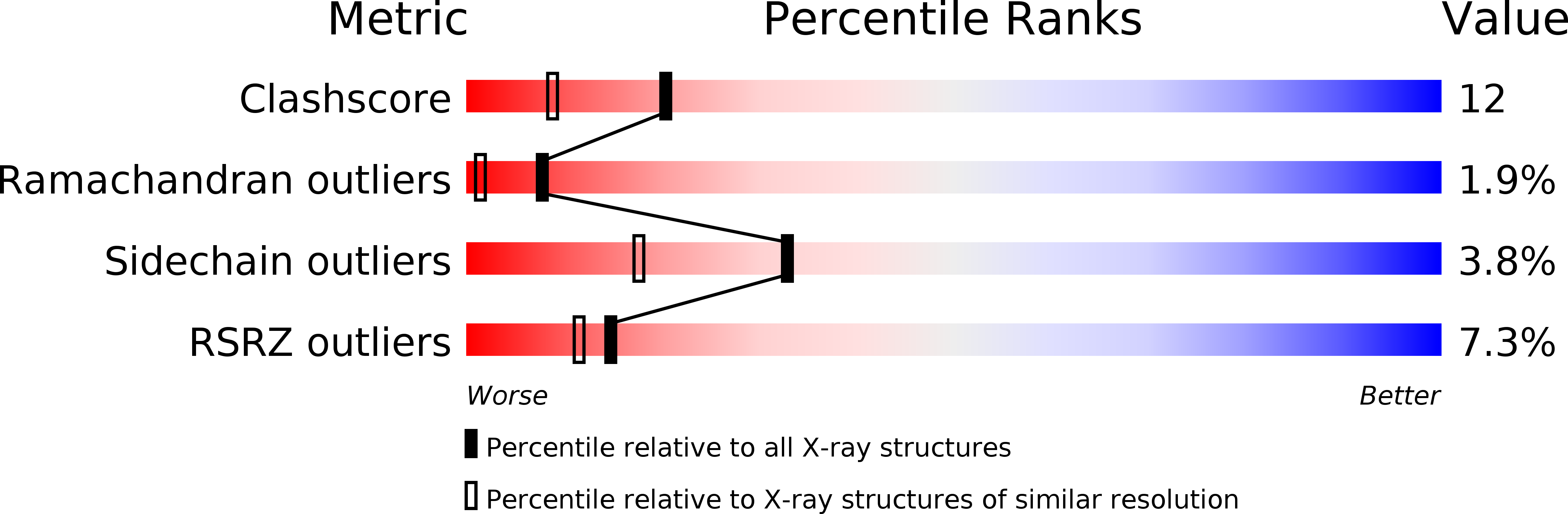
Resolution:
1.81 Å
R-Value Free:
0.26
R-Value Work:
0.23
R-Value Observed:
0.23
Space Group:
P 1