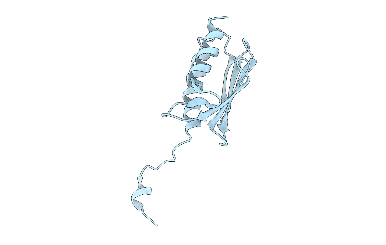
2FQL image
Deposition Date 2006-01-18
Release Date 2006-11-07
Last Version Date 2023-08-30
Entry Detail
PDB ID:
2FQL
Title:
Crystal structure of trimeric frataxin from the yeast Saccharomyces cerevisiae
Biological Source:
Source Organism:
Host Organism:
Method Details:
Experimental Method:
Resolution:
3.01 Å
R-Value Free:
0.30
R-Value Work:
0.28
R-Value Observed:
0.28
Space Group:
I 21 3
Macromolecular Entities
Polymer Type:polypeptide(L)
Molecule:Frataxin homolog, mitochondrial
Gene (Uniprot):YFH1
Mutations:Y73A
Chain IDs:A
Chain Length:123
Number of Molecules:1
Biological Source:Saccharomyces cerevisiae
Ligand Molecules
Primary Citation
The structures of frataxin oligomers reveal the mechanism for the delivery and detoxification of iron.
Structure 14 1535 1546 (2006)
PMID: 17027502 DOI: 10.1016/j.str.2006.08.010

Abstact

Defects in the mitochondrial protein frataxin are responsible for Friedreich ataxia, a neurodegenerative and cardiac disease that affects 1:40,000 children. Here, we present the crystal structures of the iron-free and iron-loaded frataxin trimers, and a single-particle electron microscopy reconstruction of a 24 subunit oligomer. The structures reveal fundamental aspects of the frataxin mechanism. The trimer has a central channel in which one atom of iron binds. Two conformations of the channel with different metal-binding affinities suggest that a gating mechanism controls whether the bound iron is delivered to other proteins or transferred to detoxification sites. The trimer constitutes the basic structural unit of the 24 subunit oligomer. The architecture of this oligomer and several features of the trimer structure demonstrate striking similarities to the iron-storage protein ferritin. The data reveal how stepwise assembly provides frataxin with the structural flexibility to perform two functions: metal delivery and detoxification.

Legend

Protein

Chemical

Disease

Primary Citation of related structures