2FOO image
Entry Detail
PDB ID:
2FOO
Keywords:
Title:
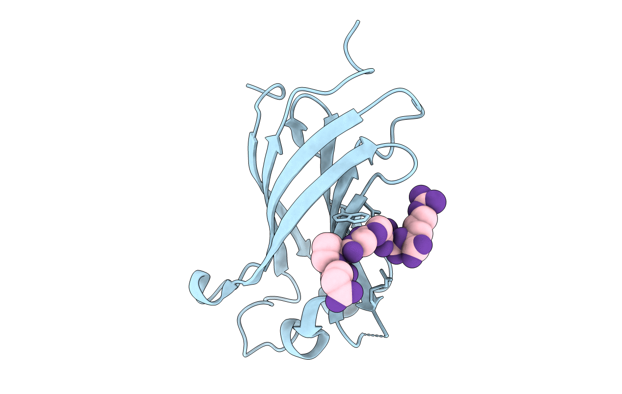
The Crystal Structure of the N-terminal domain of HAUSP/USP7 complexed with p53 peptide 359-362
Biological Source:
Source Organism:
PDB Version:
Deposition Date:
2006-01-13
Release Date:
2006-02-14
Method Details:
Experimental Method:
Resolution:
2.20 Å
R-Value Free:
0.24
R-Value Work:
0.20
R-Value Observed:
0.20
Space Group:
P 41
Macromolecular Entities
Polymer Type:polypeptide(L)
Description:Ubiquitin carboxyl-terminal hydrolase 7
Chain IDs:A
Chain Length:155
Number of Molecules:1
Biological Source:Homo sapiens
Primary Citation
Molecular recognition of p53 and MDM2 by USP7/HAUSP
Nat.Struct.Mol.Biol. 13 285 291 (2006)
PMID: 16474402 DOI: 10.1038/nsmb1067

Abstact

The ubiquitin-specific protease, USP7, has key roles in the p53 pathway whereby it stabilizes both p53 and MDM2. We show that the N-terminal domain of USP7 binds two closely spaced 4-residue sites in both p53 and MDM2, falling between p53 residues 359-367 and MDM2 residues 147-159. Cocrystal structures with USP7 were determined for both p53 peptides and for one MDM2 peptide. These peptides bind the same surface of USP7 as Epstein-Barr nuclear antigen-1, explaining the competitive nature of the interactions. The structures and mutagenesis data indicate a preference for a P/AXXS motif in peptides that bind USP7. Contacts made by serine are identical and crucial for all peptides, and Trp165 in the peptide-binding pocket of USP7 is also crucial. These results help to elucidate the mechanism of substrate recognition by USP7 and the regulation of the p53 pathway.

Legend

Protein

Chemical

Disease

Primary Citation of related structures