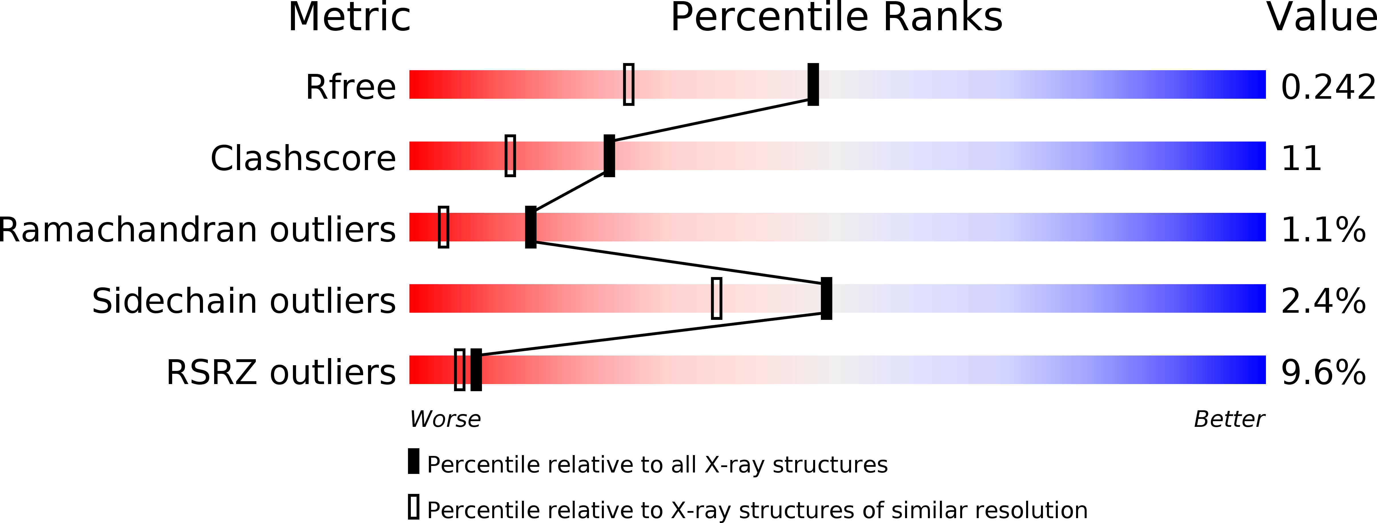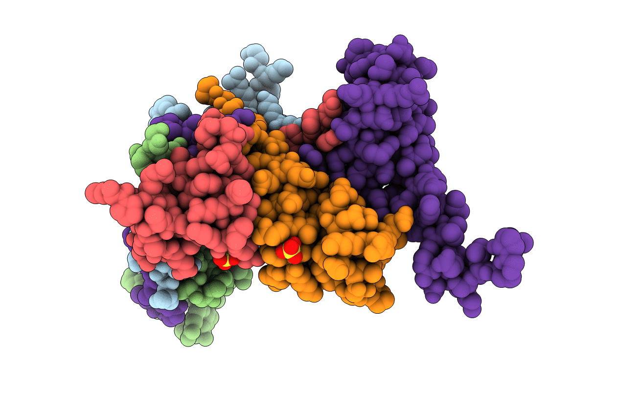
2FMM image
Deposition Date 2006-01-09
Release Date 2006-05-23
Last Version Date 2024-02-14
Entry Detail
PDB ID:
2FMM
Keywords:
Title:
Crystal Structure of EMSY-HP1 complex
Biological Source:
Source Organism:
Homo sapiens (Taxon ID: 9606)
Method Details:
Experimental Method:
Resolution:
1.80 Å
R-Value Free:
0.24
R-Value Work:
0.21
R-Value Observed:
0.21
Space Group:
P 21 21 2
Macromolecular Entities
Polymer Type:polypeptide(L)
Molecule:Chromobox protein homolog 1
Gene (Uniprot):CBX1
Chain IDs:B (auth: A), C (auth: B), D (auth: C), E (auth: D)
Chain Length:74
Number of Molecules:4
Biological Source:Homo sapiens
Polymer Type:polypeptide(L)
Molecule:Protein EMSY
Gene (Uniprot):EMSY
Chain IDs:A (auth: E)
Chain Length:133
Number of Molecules:1
Biological Source:Homo sapiens
Ligand Molecules
Primary Citation
Crystal structure of the HP1-EMSY complex reveals an unusual mode of HP1 binding.
Structure 14 703 712 (2006)
PMID: 16615912 DOI: 10.1016/j.str.2006.01.007

Abstact

Heterochromatin protein-1 (HP1) plays an essential role in both the assembly of higher-order chromatin structure and epigenetic inheritance. The C-terminal chromo shadow domain (CSD) of HP1 is responsible for homodimerization and interaction with a number of chromatin-associated nonhistone proteins, including EMSY, which is a BRCA2-interacting protein that has been implicated in the development of breast and ovarian cancer. We have determined the crystal structure of the HP1beta CSD in complex with the N-terminal domain of EMSY at 1.8 A resolution. Surprisingly, the structure reveals that EMSY is bound by two HP1 CSD homodimers, and the binding sequences differ from the consensus HP1 binding motif PXVXL. This structural information expands our understanding of HP1 binding specificity and provides insights into interactions between HP1 homodimers that are likely to be important for heterochromatin formation.

Legend

Protein

Chemical

Disease

Primary Citation of related structures