2FL5 image
Deposition Date 2006-01-05
Release Date 2006-02-21
Last Version Date 2024-10-16
Entry Detail
PDB ID:
2FL5
Keywords:
Title:
Cofactor-containing antibodies: Crystal structure of the original yellow antibody
Biological Source:
Source Organism:
Homo sapiens (Taxon ID: 9606)
Method Details:
Experimental Method:
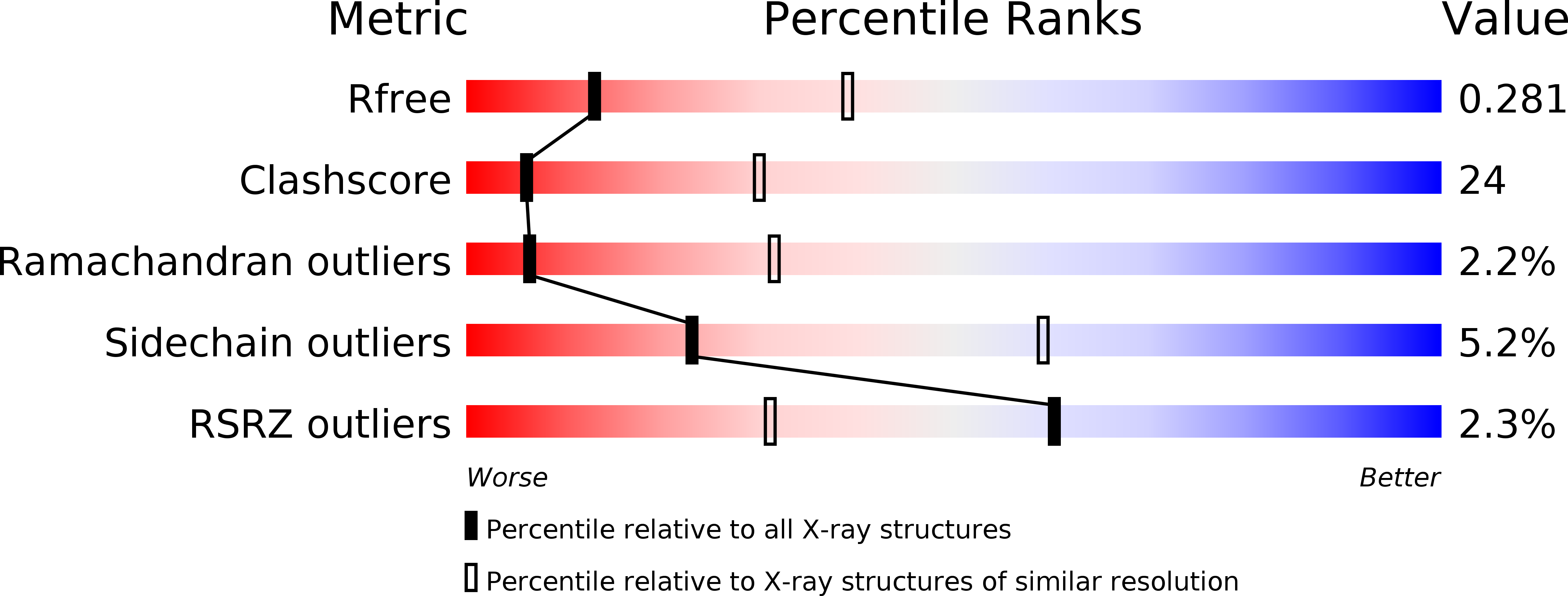
Resolution:
3.00 Å
R-Value Free:
0.29
R-Value Work:
0.24
R-Value Observed:
0.24
Space Group:
P 21 21 21
Macromolecular Entities
Polymer Type:polypeptide(L)
Molecule:Immunoglobulin Igg1 Heavy chain
Chain IDs:B (auth: H), D (auth: B), F (auth: D), H (auth: F)
Chain Length:220
Number of Molecules:4
Biological Source:Homo sapiens
Structures with similar UniProt ID
Protein Blast
Polymer Type:polypeptide(L)
Molecule:Immunoglobulin Igg1 Lambda Light Chain
Gene (Uniprot):IGL@
Chain IDs:A (auth: L), C (auth: A), E (auth: C), G (auth: E)
Chain Length:209
Number of Molecules:4
Biological Source:Homo sapiens
Ligand Molecules
Primary Citation
Cofactor-containing antibodies: Crystal structure of the original yellow antibody.
Proc.Natl.Acad.Sci.Usa 103 3581 3585 (2006)
PMID: 16537445 DOI: 10.1073/pnas.0600251103

Abstact

Antibodies are generally thought to be a class of proteins that function without the use of cofactors. However, it is not widely appreciated that antibodies are believed to be the major carrier protein in human circulation for the important riboflavin cofactor that is involved in a host of biological phenomena. A further link between riboflavin and antibodies was discovered 30 years ago when a bright-yellow antibody, IgG(GAR), was purified from a patient with multiple myeloma who had turned yellow during the course of her disease. It was subsequently shown that the yellow color of this antibody was due to riboflavin binding. However, it was not known how and where riboflavin was bound to this antibody. We now report the crystal structure of this historically important IgG(GAR) Fab at 3.0-A resolution. The riboflavin is located in the antigen-combining site with its isoalloxazine ring stacked between the parallel aromatic moieties of TyrH33, PheH58, and TyrH100A. Together with additional hydrogen bonds, these interactions reveal the structural basis for high-affinity riboflavin binding. The ligand specificity of IgG(GAR) is compared with another riboflavin-binding antibody, IgG(DOT), which was purified from a second patient with multiple myeloma. The crystal structure of IgG(GAR) provides a starting point for attempts to understand the physiological relevance and chemical functions of cofactor-containing antibodies.

Legend

Protein

Chemical

Disease

Primary Citation of related structures
Feedback Form
Name
Email
Institute
Feedback