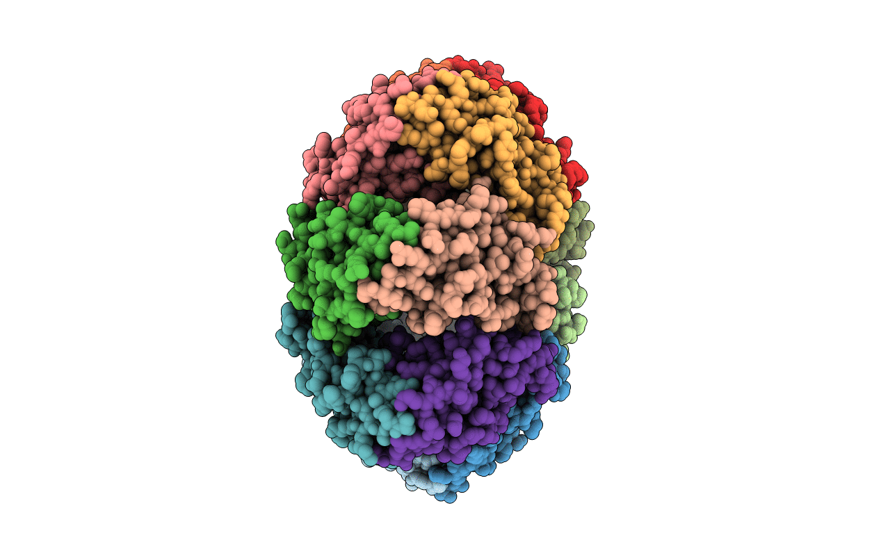
2FKD image
Deposition Date 2006-01-04
Release Date 2006-06-13
Last Version Date 2024-02-14
Entry Detail
PDB ID:
2FKD
Title:
Crystal Structure of the C-terminal domain of Bacteriophage 186 repressor
Biological Source:
Source Organism:
Host Organism:
Method Details:
Experimental Method:
Resolution:
2.70 Å
R-Value Free:
0.29
R-Value Work:
0.24
R-Value Observed:
0.24
Space Group:
P 21 21 21
Macromolecular Entities
Polymer Type:polypeptide(L)
Molecule:Repressor protein CI
Gene (Uniprot):CI
Chain IDs:A, B, C, D, E, F, G, H, I, J, K, L, M, N
Chain Length:110
Number of Molecules:14
Biological Source:Enterobacteria phage 186
Ligand Molecules
Primary Citation
The structural basis of cooperative regulation at an alternate genetic switch.
Mol.Cell 21 605 615 (2006)
PMID: 16507359 DOI: 10.1016/j.molcel.2006.01.019

Abstact

Bacteriophage lambda is a paradigm for understanding the role of cooperativity in gene regulation. Comparison of the regulatory regions of lambda and the unrelated temperate bacteriophage 186 provides insight into alternate ways to assemble functional genetic switches. The structure of the C-terminal domain of the 186 repressor, determined at 2.7 A resolution, reveals an unusual heptamer of dimers, consistent with presented genetic studies. In addition, the structure of a cooperativity mutant of the full-length 186 repressor, identified by genetic screens, was solved to 1.95 A resolution. These structures provide a molecular basis for understanding lysogenic regulation in 186. Whereas the overall fold of the 186 and lambda repressor monomers is remarkably similar, the way the two repressors cooperatively assemble is quite different and explains in part the differences in their regulatory activity.

Legend

Protein

Chemical

Disease

Primary Citation of related structures