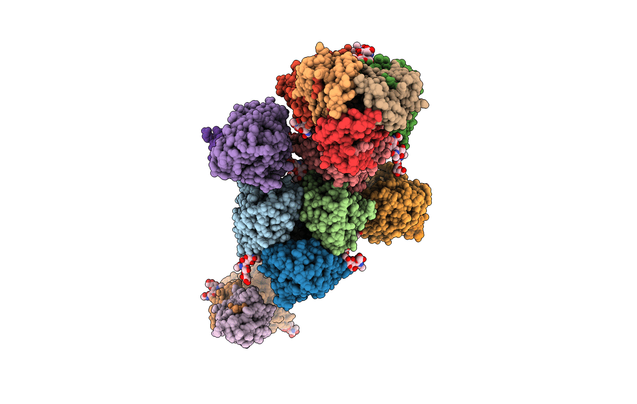
2FK0 image
Entry Detail
PDB ID:
2FK0
Keywords:
Title:
Crystal Structure of a H5N1 influenza virus hemagglutinin.
Biological Source:
PDB Version:
Deposition Date:
2006-01-03
Release Date:
2006-05-02
Method Details:
Experimental Method:
Resolution:
2.95 Å
R-Value Free:
0.31
R-Value Work:
0.26
R-Value Observed:
0.26
Space Group:
P 3
Macromolecular Entities
Polymer Type:polypeptide(L)
Description:hemagglutinin
Chain IDs:A, C, E, G, I, K, M, O, Q
Chain Length:334
Number of Molecules:9
Biological Source:Influenza A virus (A/Viet Nam/1203/2004(H5N1))
Polymer Type:polypeptide(L)
Description:hemagglutinin
Chain IDs:B, D, F, H, J, L, N, P, R
Chain Length:181
Number of Molecules:9
Biological Source:Influenza A virus (A/Viet Nam/1203/2004(H5N1))
Modified Residue
Compound ID Chain ID Parent Comp ID Details 2D Image
ASN A ASN GLYCOSYLATION SITE
Ligand Molecules
Primary Citation
Structure and Receptor Specificity of the Hemagglutinin from an H5N1 Influenza Virus.
Science 312 404 410 (2006)
PMID: 16543414 DOI: 10.1126/science.1124513

Abstact

The hemagglutinin (HA) structure at 2.9 angstrom resolution, from a highly pathogenic Vietnamese H5N1 influenza virus, is more related to the 1918 and other human H1 HAs than to a 1997 duck H5 HA. Glycan microarray analysis of this Viet04 HA reveals an avian alpha2-3 sialic acid receptor binding preference. Introduction of mutations that can convert H1 serotype HAs to human alpha2-6 receptor specificity only enhanced or reduced affinity for avian-type receptors. However, mutations that can convert avian H2 and H3 HAs to human receptor specificity, when inserted onto the Viet04 H5 HA framework, permitted binding to a natural human alpha2-6 glycan, which suggests a path for this H5N1 virus to gain a foothold in the human population.

Legend

Protein

Chemical

Disease

Primary Citation of related structures