2FJY image
Deposition Date 2006-01-03
Release Date 2006-01-17
Last Version Date 2024-10-09
Entry Detail
PDB ID:
2FJY
Title:
Crystal Structure of B-form Bombyx mori Pheromone Binding Protein
Biological Source:
Source Organism:
Bombyx mori (Taxon ID: 7091)
Host Organism:
Method Details:
Experimental Method:
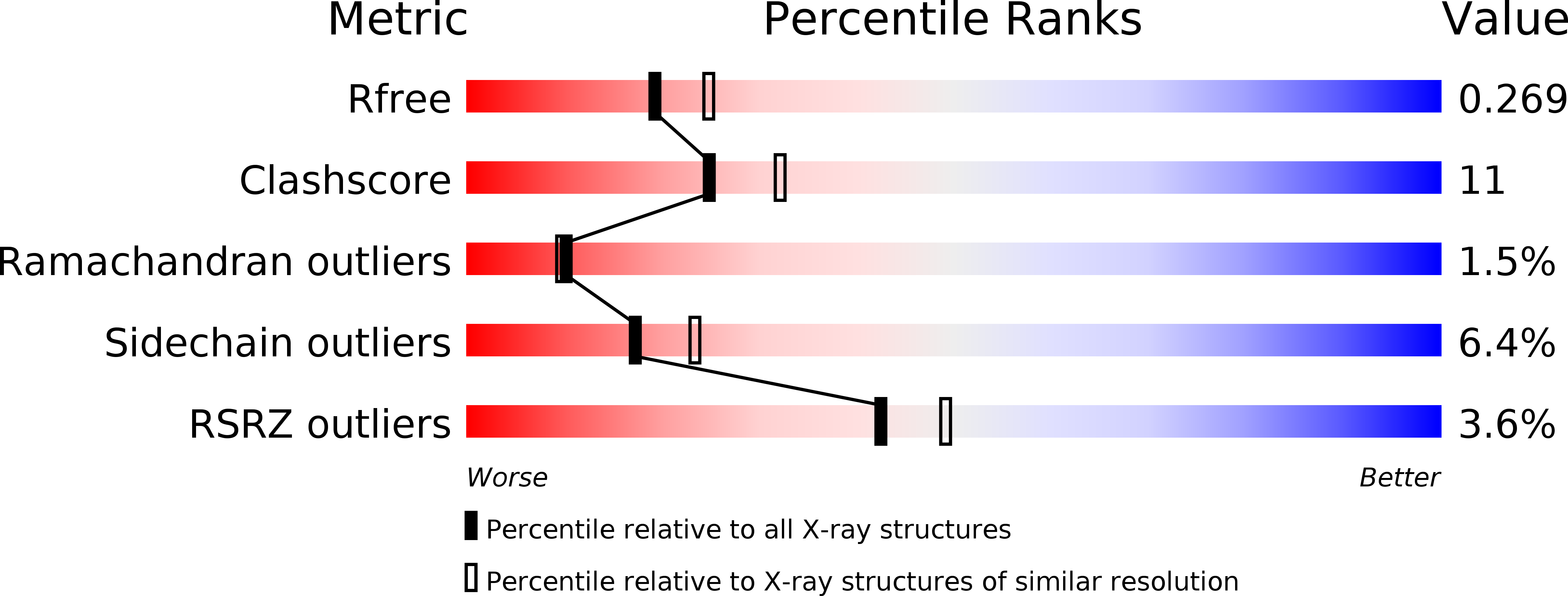
Resolution:
2.30 Å
R-Value Free:
0.27
R-Value Work:
0.22
R-Value Observed:
0.22
Space Group:
P 21 21 21
Macromolecular Entities
Polymer Type:polypeptide(L)
Molecule:Pheromone-binding protein
Chain IDs:A, B
Chain Length:142
Number of Molecules:2
Biological Source:Bombyx mori
Primary Citation
Coil-to-helix transition and ligand release of Bombyx mori pheromone-binding protein.
Biochem.Biophys.Res.Commun. 335 1044 1050 (2005)
PMID: 16111659 DOI: 10.1016/j.bbrc.2005.07.176

Abstact

The transport of hydrophobic insect pheromones through the aqueous medium surrounding their receptors is assisted by pheromone-binding proteins (PBPs). The protein from the silkworm moth Bombyx mori, BmorPBP, exhibits a pH-dependent conformational change postulated to trigger the release of the pheromone bombykol to its receptor. At low pH, an alpha-helix occupies the same binding pocket that houses the pheromone in the BmorPBP-bombykol complex at high pH. We have determined the crystal structure of apo BmorPBP at a resolution of 2.3 angstroms and pH 7.5, which has surprisingly a structure similar to the A-form. These data suggest that BmorPBP undergoes a ligand-dependent conformational change in addition to the previously described pH-dependent conformational change. Analysis of the alpha-helix occupying the binding pocket reveals an amphipathic helix with three acidic residues along one face that are conserved among lepidopteran PBPs and may be involved in a conformational transition of BmorPBP at the receptor membrane.

Legend

Protein

Chemical

Disease

Primary Citation of related structures