Deposition Date
2006-01-02
Release Date
2006-10-31
Last Version Date
2024-02-14
Entry Detail
PDB ID:
2FJ9
Keywords:
Title:
High resolution crystal structure of the unliganded human ACBP
Biological Source:
Source Organism(s):
Homo sapiens (Taxon ID: 9606)
Expression System(s):
Method Details:
Experimental Method:
Resolution:
1.60 Å
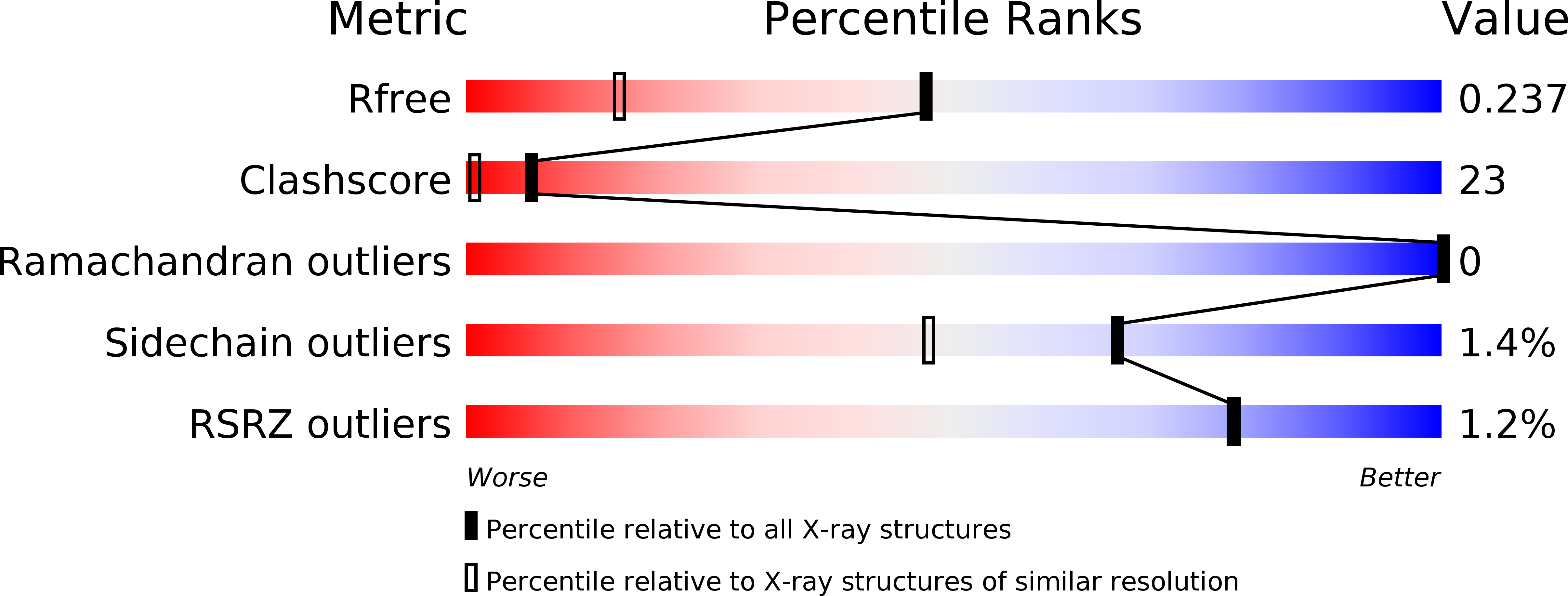
R-Value Free:
0.22
R-Value Work:
0.19
R-Value Observed:
0.19
Space Group:
P 65 2 2