2FIV image
Deposition Date 1997-07-21
Release Date 1997-11-12
Last Version Date 2023-08-09
Entry Detail
PDB ID:
2FIV
Title:
Crystal structure of feline immunodeficiency virus protease complexed with a substrate
Biological Source:
Source Organism:
Host Organism:
Method Details:
Experimental Method:
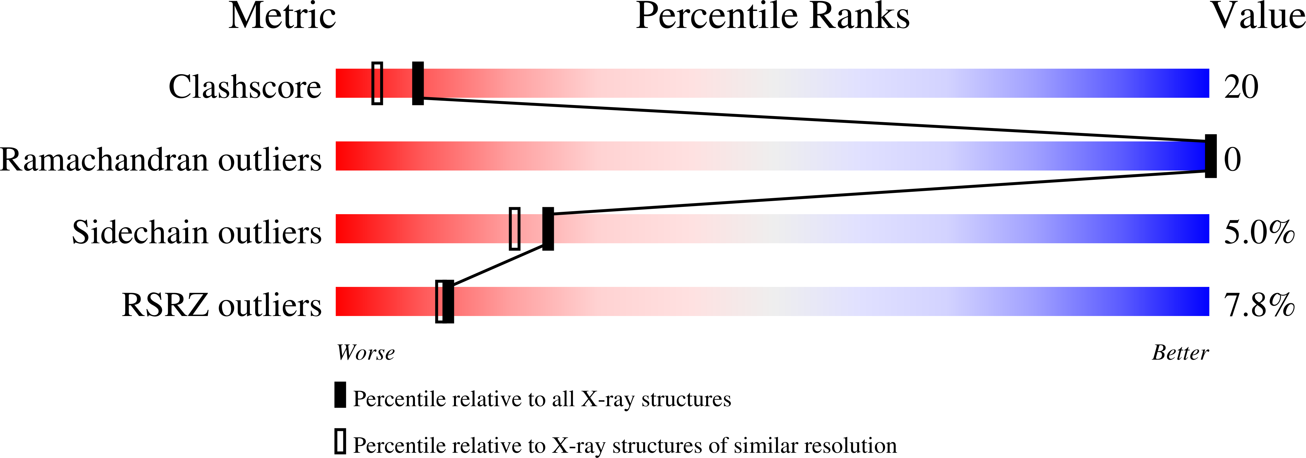
Resolution:
2.00 Å
R-Value Work:
0.16
R-Value Observed:
0.16
Space Group:
P 31
Macromolecular Entities
Structures with similar UniProt ID
Protein Blast
Polymer Type:polypeptide(L)
Molecule:FELINE IMMUNODEFICIENCY VIRUS PROTEASE
Gene (Uniprot):pol
Mutagens:D30N
Chain IDs:A, B
Chain Length:116
Number of Molecules:2
Biological Source:Feline immunodeficiency virus
Modified Residue
Compound ID Chain ID Parent Comp ID Details 2D Image
ALN C ALA NAPHTHALEN-2-YL-3-ALANINE
Ligand Molecules
Peptide-like Molecules
PRD_000427
Primary Citation
Crystal structures of the inactive D30N mutant of feline immunodeficiency virus protease complexed with a substrate and an inhibitor.
Biochemistry 36 10696 10708 (1997)
PMID: 9271500 DOI: 10.1021/bi9707436

Abstact

Crystal structures of complexes of a D30N mutant of feline immunodeficiency virus protease (FIV PR) complexed with a statine-based inhibitor (LP-149), as well as with a substrate based on a modification of this inhibitor (LP-149S), have been solved and refined at resolutions of 2.0 and 1.85 A, respectively. Both the inhibitor and the substrate are bound in the active site of the mutant protease in a similar mode, which also resembles the mode of binding of LP-149 to the native protease. The carbonyl oxygen of the scissile bond in the substrate is not hydrated and is located within the distance of a hydrogen bond to an amido nitrogen atom from one of the two asparagines in the active site of the enzyme. The nitrogen atom of the scissile bond is 3.25 A from the conserved water molecule (Wat301). A model of a tetrahedral intermediate bound to the active site of the native enzyme was built by considering the interactions observed in all three crystal structures of FIV PR. Molecular dynamics simulations of this model bound to native wild-type FIV PR were carried out, to investigate the final stages of the catalytic mechanism of aspartic proteases.

Legend

Protein

Chemical

Disease

Primary Citation of related structures