
Deposition Date
2005-12-29
Release Date
2006-02-07
Last Version Date
2023-08-30
Entry Detail
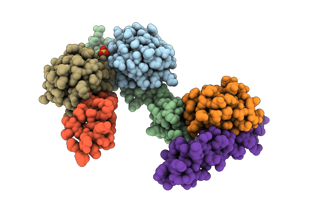
PDB ID:
2FIF
Keywords:
Title:
Crystal Structure of a Bovine Rabex-5 fragment complexed with ubiquitin
Biological Source:
Source Organism(s):
Bos taurus (Taxon ID: 9913)
Expression System(s):
Method Details:
Experimental Method:
Resolution:
2.49 Å
R-Value Free:
0.26
R-Value Work:
0.22
R-Value Observed:
0.23
Space Group:
C 1 2 1
