2FFL image
Deposition Date 2005-12-19
Release Date 2006-01-24
Last Version Date 2024-02-14
Entry Detail
PDB ID:
2FFL
Title:
Crystal Structure of Dicer from Giardia intestinalis
Biological Source:
Source Organism:
Host Organism:
Method Details:
Experimental Method:
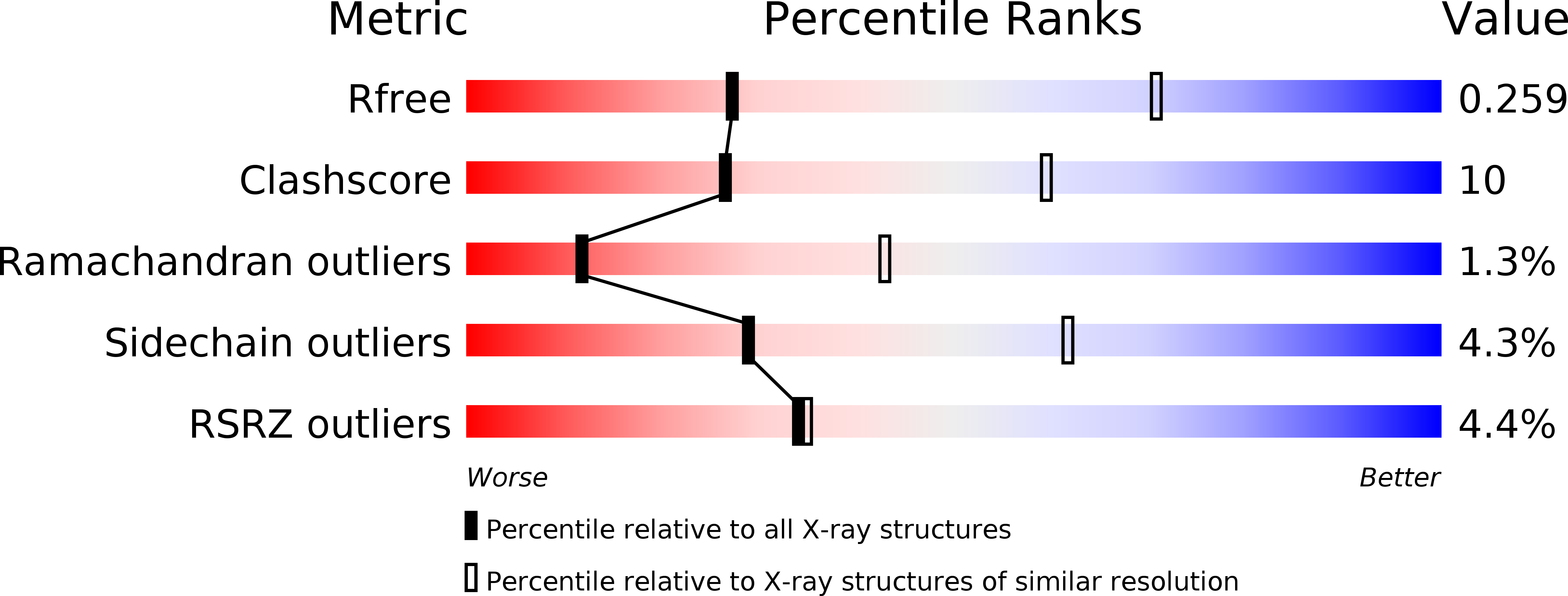
Resolution:
3.33 Å
R-Value Free:
0.27
R-Value Work:
0.24
R-Value Observed:
0.24
Space Group:
P 21 21 2
Macromolecular Entities
Polymer Type:polypeptide(L)
Molecule:Dicer
Mutations:N-terminal GA residues
Chain IDs:A, B, C, D
Chain Length:756
Number of Molecules:4
Biological Source:Giardia intestinalis
Ligand Molecules
Primary Citation
Structural Basis of Double-Stranded RNA Processing by Dicer
Science 311 195 198 (2006)
PMID: 16410517 DOI: 10.1126/science.1121638

Abstact

The specialized ribonuclease Dicer initiates RNA interference by cleaving double-stranded RNA (dsRNA) substrates into small fragments about 25 nucleotides in length. In the crystal structure of an intact Dicer enzyme, the PAZ domain, a module that binds the end of dsRNA, is separated from the two catalytic ribonuclease III (RNase III) domains by a flat, positively charged surface. The 65 angstrom distance between the PAZ and RNase III domains matches the length spanned by 25 base pairs of RNA. Thus, Dicer itself is a molecular ruler that recognizes dsRNA and cleaves a specified distance from the helical end.

Legend

Protein

Chemical

Disease

Primary Citation of related structures