2FF6 image
Deposition Date 2005-12-19
Release Date 2006-03-21
Last Version Date 2024-11-06
Entry Detail
PDB ID:
2FF6
Title:
Crystal structure of Gelsolin domain 1:ciboulot domain 2 hybrid in complex with actin
Biological Source:
Source Organism:
Host Organism:
Method Details:
Experimental Method:
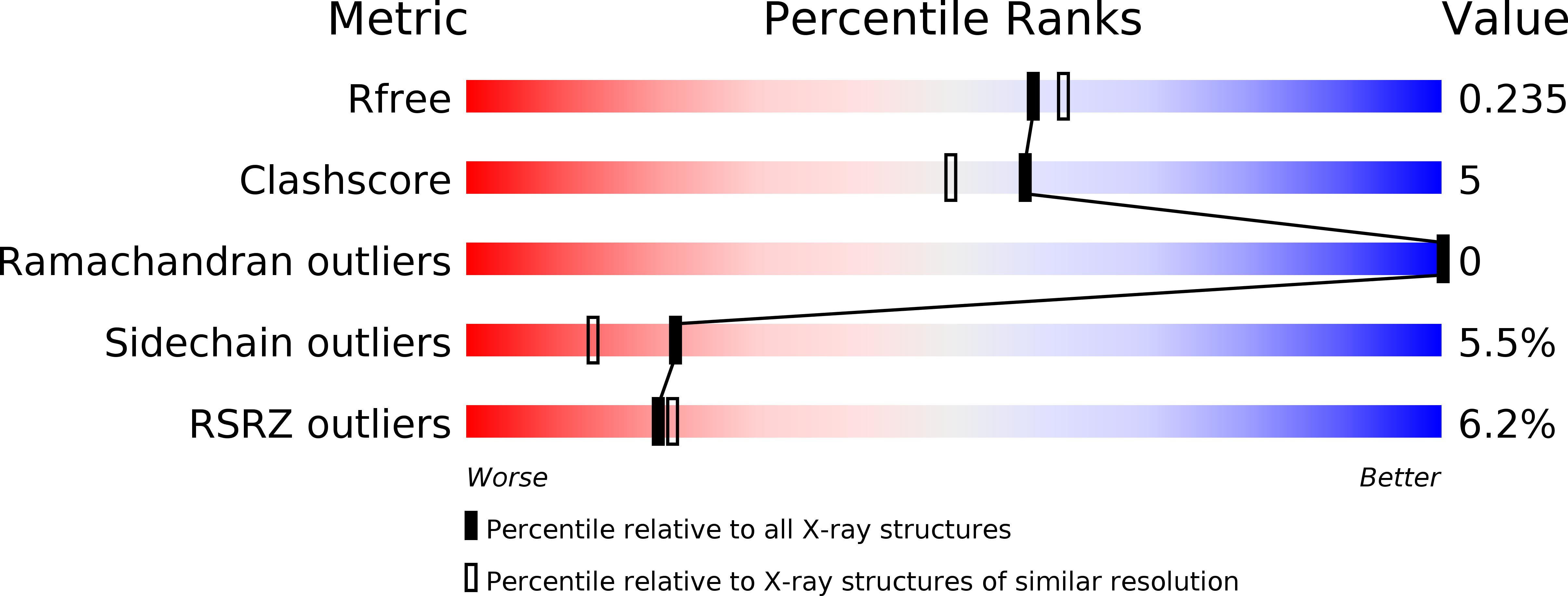
Resolution:
2.05 Å
R-Value Free:
0.23
R-Value Work:
0.20
R-Value Observed:
0.20
Space Group:
P 1 21 1
Macromolecular Entities
Polymer Type:polypeptide(L)
Molecule:Actin, alpha skeletal muscle
Gene (Uniprot):ACTA1
Chain IDs:C (auth: A)
Chain Length:375
Number of Molecules:1
Biological Source:Oryctolagus cuniculus
Polymer Type:polypeptide(L)
Molecule:Gelsolin
Gene (Uniprot):GSN
Chain IDs:A (auth: G)
Chain Length:150
Number of Molecules:1
Biological Source:Homo sapiens
Polymer Type:polypeptide(L)
Molecule:CG4944-PC, isoform C
Gene (Uniprot):cib
Chain IDs:B (auth: H)
Chain Length:26
Number of Molecules:1
Biological Source:Drosophila melanogaster
Primary Citation
The Structural Basis of Actin Interaction with Multiple WH2/beta-Thymosin Motif-Containing Proteins
Structure 14 469 476 (2006)
PMID: 16531231 DOI: 10.1016/j.str.2005.12.011

Abstact

Participation of actin in cellular processes relies on the dynamics of filament assembly. Filament elongation is fed by monomeric actin in complex with either profilin or a Wiscott-Aldrich syndrome protein (WASP) homology domain 2 (WH2)/beta-thymosin (betaT) domain. WH2/betaT motif repetition (typified by ciboulot) or combination with nonrelated domains (as found in N-WASP) results in proteins that yield their actin to filament elongation. Here, we report the crystal structures of actin bound hybrid proteins, constructed between gelsolin and WH2/betaT domains from ciboulot or N-WASP. We observe the C-terminal half of ciboulot domain 2 bound to actin. In solution, we show that cibolout domains 2 and 3 bind to both G- and F-actin, and that whole ciboulot forms a complex with two actin monomers. In contrast, the analogous portion of N-WASP WH2 domain 2 is detached from actin, indicating that the C-terminal halves of the betaT and WH2 motifs are not functionally analogous.

Legend

Protein

Chemical

Disease

Primary Citation of related structures