Deposition Date
2005-12-18
Release Date
2006-05-23
Last Version Date
2023-09-20
Entry Detail
PDB ID:
2FF2
Keywords:
Title:
Crystal structure of Trypanosoma vivax nucleoside hydrolase co-crystallized with ImmucillinH
Biological Source:
Source Organism(s):
Trypanosoma vivax (Taxon ID: 5699)
Expression System(s):
Method Details:
Experimental Method:
Resolution:
2.20 Å
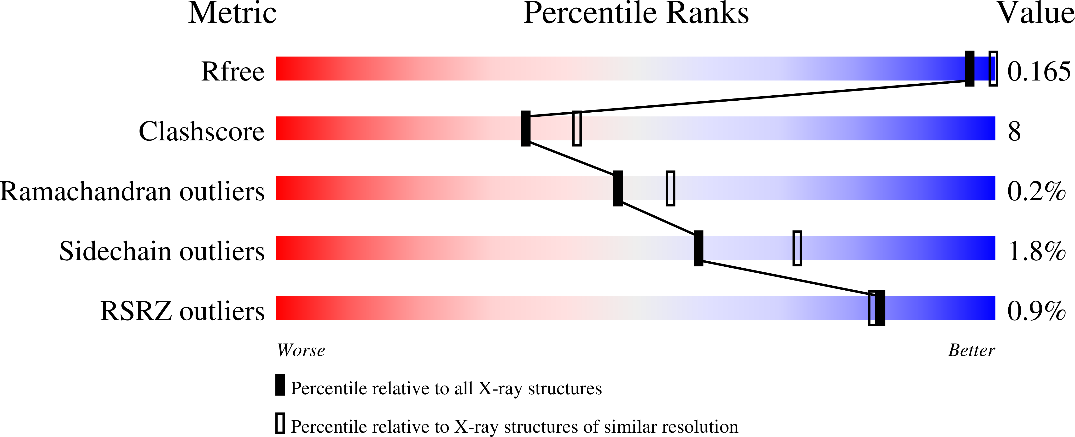
R-Value Free:
0.22
R-Value Work:
0.16
R-Value Observed:
0.16
Space Group:
P 1 21 1