2FE4 image
Deposition Date 2005-12-15
Release Date 2006-07-04
Last Version Date 2024-02-14
Entry Detail
PDB ID:
2FE4
Keywords:
Title:
The crystal structure of human neuronal Rab6B in its inactive GDP-bound form
Biological Source:
Source Organism:
Homo sapiens (Taxon ID: 9606)
Method Details:
Experimental Method:
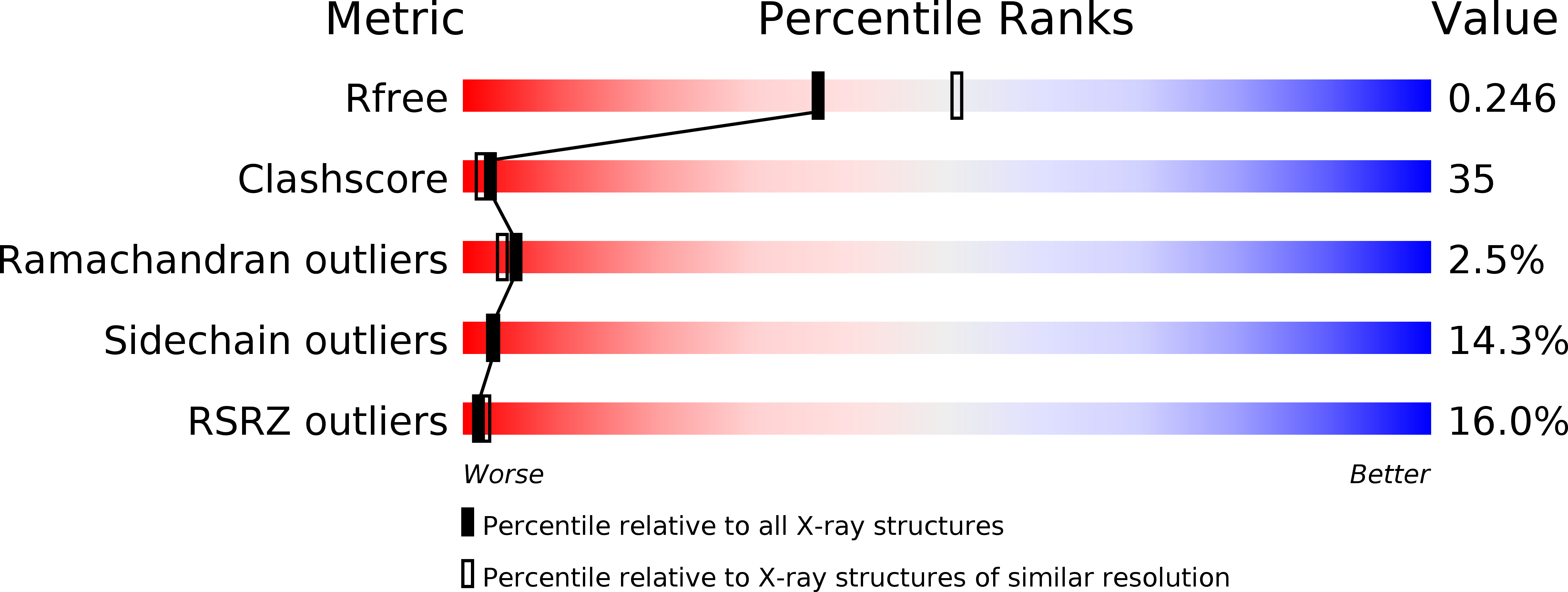
Resolution:
2.30 Å
R-Value Free:
0.25
R-Value Work:
0.22
R-Value Observed:
0.22
Space Group:
P 21 21 21
Macromolecular Entities
Structures with similar UniProt ID
Protein Blast
Polymer Type:polypeptide(L)
Molecule:Ras-related protein Rab-6B
Gene (Uniprot):RAB6B
Chain IDs:A
Chain Length:171
Number of Molecules:1
Biological Source:Homo sapiens
Primary Citation
The structure of human neuronal Rab6B in the active and inactive form.
Acta Crystallogr.,Sect.D 62 725 733 (2006)
PMID: 16790928 DOI: 10.1107/S0907444906015319

Abstact

The Rab small G-protein family plays important roles in eukaryotes as regulators of vesicle traffic. In Rab proteins, the hydrolysis of GTP to GDP is coupled with association with and dissociation from membranes. Conformational changes related to their different nucleotide states determine their effector specificity. The crystal structure of human neuronal Rab6B was solved in its 'inactive' (with bound MgGDP) and 'active' (MgGTPgammaS-bound) forms to 2.3 and 1.8 A, respectively. Both crystallized in space group P2(1)2(1)2(1), with similar unit-cell parameters, allowing the comparison of both structures without packing artifacts. Conformational changes between the inactive GDP and active GTP-like state are observed mainly in the switch I and switch II regions, confirming their role as a molecular switch. Compared with other Rab proteins, additional changes are observed in the Rab6 subfamily-specific RabSF3 region that might contribute to the specificity of Rab6 for its different effector proteins.

Legend

Protein

Chemical

Disease

Primary Citation of related structures