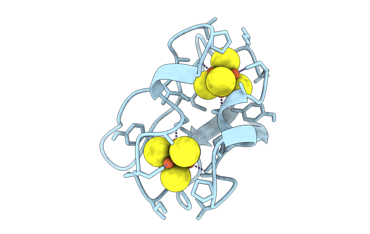
2FDN image
Deposition Date 1997-10-01
Release Date 1998-04-08
Last Version Date 2024-02-14
Entry Detail
PDB ID:
2FDN
Title:
2[4FE-4S] FERREDOXIN FROM CLOSTRIDIUM ACIDI-URICI
Biological Source:
Source Organism:
Method Details:
Experimental Method:
Resolution:
0.94 Å
R-Value Observed:
0.10
Space Group:
P 43 21 2
Macromolecular Entities
Structures with similar UniProt ID
Protein Blast
Polymer Type:polypeptide(L)
Molecule:FERREDOXIN
Gene (Uniprot):fdxA
Chain IDs:A
Chain Length:55
Number of Molecules:1
Biological Source:Clostridium acidurici
Ligand Molecules
Primary Citation
Atomic resolution (0.94 A) structure of Clostridium acidurici ferredoxin. Detailed geometry of [4Fe-4S] clusters in a protein.
Biochemistry 36 16065 16073 (1997)
PMID: 9405040 DOI: 10.1021/bi972155y

Abstact

The crystal structure of the 2[4Fe-4S] ferredoxin from Clostridium acidurici has been solved using X-ray diffraction data extending to atomic resolution, 0.94 A, recorded at 100 K. The model was refined with anisotropic representation of atomic displacement parameters for all non-hydrogen atoms and with hydrogens riding on their parent atoms. Stereochemical restraints were applied to the protein chain but not to the iron-sulfur clusters. The final R factor is 10.03 % for all data. Inversion of the final least-squares matrix allowed direct estimation of the errors of individual parameters. The estimated errors in positions for protein main chain atoms are below 0.02 A and about 0.003 A for the heavier [4Fe-4S] cluster atoms. Significant differences between the stereochemistry of the two clusters and distortion of both of them from ideal Td tetrahedral symmetry can be defined in detail at this level of accuracy. Regions of alternative conformations include not only protein side chains but also two regions of the main chain. One such region is the loop of residues 25-29, which was highly disordered in the room temperature structure.

Legend

Protein

Chemical

Disease

Primary Citation of related structures