2FDF image
Entry Detail
PDB ID:
2FDF
Title:
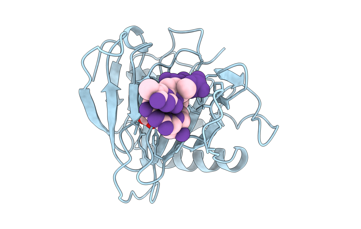
Crystal Structure of AlkB in complex with Co(II), 2-oxoglutarate, and methylated trinucleotide T-meA-T
Biological Source:
Source Organism:
PDB Version:
Deposition Date:
2005-12-13
Release Date:
2006-02-21
Method Details:
Experimental Method:
Resolution:
2.10 Å
R-Value Free:
0.23
R-Value Work:
0.19
Space Group:
P 43
Macromolecular Entities
Polymer Type:polypeptide(L)
Description:Alkylated DNA repair protein alkB
Chain IDs:B (auth: A)
Chain Length:211
Number of Molecules:1
Biological Source:Escherichia coli K12
Modified Residue
Compound ID Chain ID Parent Comp ID Details 2D Image
MA7 A DA 1N-METHYLADENOSINE-5'-MONOPHOSPHATE
Primary Citation
Crystal structures of catalytic complexes of the oxidative DNA/RNA repair enzyme AlkB.
Nature 439 879 884 (2006)
PMID: 16482161 DOI: 10.1038/nature04561

Abstact

Nucleic acid damage by environmental and endogenous alkylation reagents creates lesions that are both mutagenic and cytotoxic, with the latter effect accounting for their widespread use in clinical cancer chemotherapy. Escherichia coli AlkB and the homologous human proteins ABH2 and ABH3 (refs 5, 7) promiscuously repair DNA and RNA bases damaged by S(N)2 alkylation reagents, which attach hydrocarbons to endocyclic ring nitrogen atoms (N1 of adenine and guanine and N3 of thymine and cytosine). Although the role of AlkB in DNA repair has long been established based on phenotypic studies, its exact biochemical activity was only elucidated recently after sequence profile analysis revealed it to be a member of the Fe-oxoglutarate-dependent dioxygenase superfamily. These enzymes use an Fe(II) cofactor and 2-oxoglutarate co-substrate to oxidize organic substrates. AlkB hydroxylates an alkylated nucleotide base to produce an unstable product that releases an aldehyde to regenerate the unmodified base. Here we have determined crystal structures of substrate and product complexes of E. coli AlkB at resolutions from 1.8 to 2.3 A. Whereas the Fe-2-oxoglutarate dioxygenase core matches that in other superfamily members, a unique subdomain holds a methylated trinucleotide substrate into the active site through contacts to the polynucleotide backbone. Amide hydrogen exchange studies and crystallographic analyses suggest that this substrate-binding 'lid' is conformationally flexible, which may enable docking of diverse alkylated nucleotide substrates in optimal catalytic geometry. Different crystal structures show open and closed states of a tunnel putatively gating O2 diffusion into the active site. Exposing crystals of the anaerobic Michaelis complex to air yields slow but substantial oxidation of 2-oxoglutarate that is inefficiently coupled to nucleotide oxidation. These observations suggest that protein dynamics modulate redox chemistry and that a hypothesized migration of the reactive oxy-ferryl ligand on the catalytic Fe ion may be impeded when the protein is constrained in the crystal lattice.

Legend

Protein

Chemical

Disease

Primary Citation of related structures