Deposition Date
2005-12-13
Release Date
2006-02-07
Last Version Date
2024-11-20
Entry Detail
PDB ID:
2FDB
Keywords:
Title:
Crystal Structure of Fibroblast growth factor (FGF)8b in complex with FGF Receptor (FGFR) 2c
Biological Source:
Source Organism(s):
Homo sapiens (Taxon ID: 9606)
Expression System(s):
Method Details:
Experimental Method:
Resolution:
2.28 Å
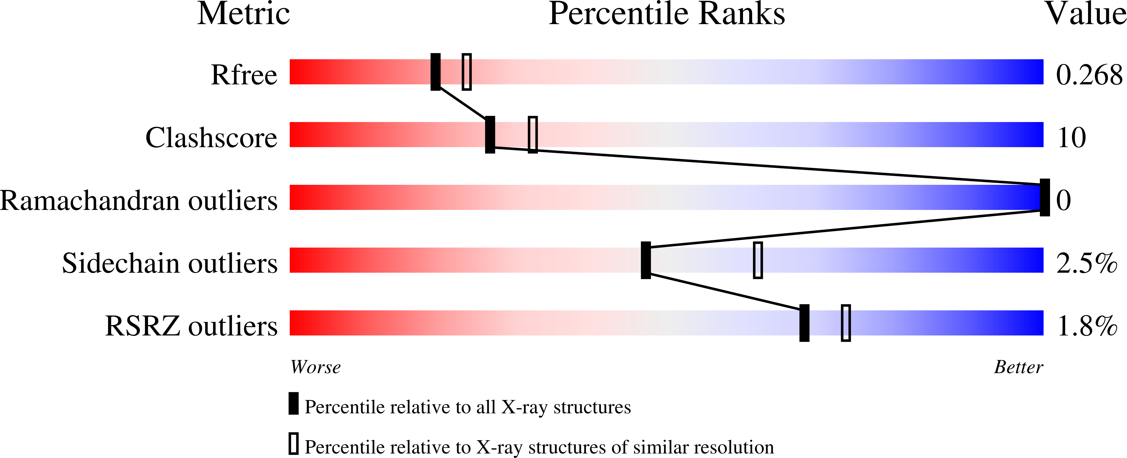
R-Value Free:
0.27
R-Value Work:
0.23
R-Value Observed:
0.23
Space Group:
C 1 2 1