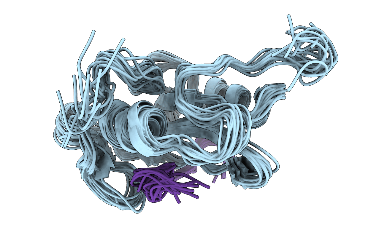
2FCI image
Deposition Date 2005-12-12
Release Date 2006-01-31
Last Version Date 2023-11-15
Entry Detail
PDB ID:
2FCI
Keywords:
Title:
Structural basis for the requirement of two phosphotyrosines in signaling mediated by Syk tyrosine kinase
Biological Source:
Source Organism:
Bos taurus (Taxon ID: 9913)
(Taxon ID: )
Method Details:
Experimental Method:
Conformers Calculated:
50
Conformers Submitted:
16
Selection Criteria:
Lack of NOE violations & correct main chain geometry
Macromolecular Entities
Polymer Type:polypeptide(L)
Molecule:C-termainl SH2 domain from phospholipase C-gamma-1 comprising residues 663-759
Gene (Uniprot):PLCG1
Chain IDs:B (auth: A)
Chain Length:105
Number of Molecules:1
Biological Source:Bos taurus
Polymer Type:polypeptide(L)
Molecule:Doubly phosphorylated peptide derived from Syk kinase comprising residues 338-350
Chain IDs:A (auth: B)
Chain Length:14
Number of Molecules:1
Biological Source:
Modified Residue
Compound ID Chain ID Parent Comp ID Details 2D Image
GMA A GLU 4-AMIDO-4-CARBAMOYL-BUTYRIC ACID
PTR A TYR O-PHOSPHOTYROSINE
Ligand Molecules
Primary Citation
Structural basis for the requirement of two phosphotyrosine residues in signaling mediated by syk tyrosine kinase
J.Mol.Biol. 356 1222 1236 (2006)
PMID: 16410013 DOI: 10.1016/j.jmb.2005.11.095

Abstact

The protein-tyrosine kinase Syk couples immune recognition receptors to multiple signal transduction pathways, including the mobilization of calcium and the activation of NFAT. The ability of Syk to regulate signaling is influenced by its phosphorylation on tyrosine residues within the linker B region. The phosphorylation of both Y342 and Y346 is necessary for optimal signaling from the B cell receptor for antigen. The SH2 domains of multiple signaling proteins share the ability to bind this doubly phosphorylated site. The NMR structure of the C-terminal SH2 domain of PLCgamma (PLCC) bound to a doubly phosphorylated Syk peptide reveals a novel mode of phosphotyrosine recognition. PLCC undergoes extensive conformational changes upon binding to form a second phosphotyrosine-binding pocket in which pY346 is largely desolvated and stabilized through electrostatic interactions. The formation of the second binding pocket is distinct from other modes of phosphotyrosine recognition in SH2-protein association. The dependence of signaling on simultaneous phosphorylation of these two tyrosine residues offers a new mechanism to fine-tune the cellular response to external stimulation.

Legend

Protein

Chemical

Disease

Primary Citation of related structures