2FBE image
Deposition Date 2005-12-09
Release Date 2006-01-10
Last Version Date 2024-11-06
Entry Detail
PDB ID:
2FBE
Title:
Crystal Structure of the PRYSPRY-domain
Biological Source:
Source Organism:
Homo sapiens (Taxon ID: 9606)
Method Details:
Experimental Method:
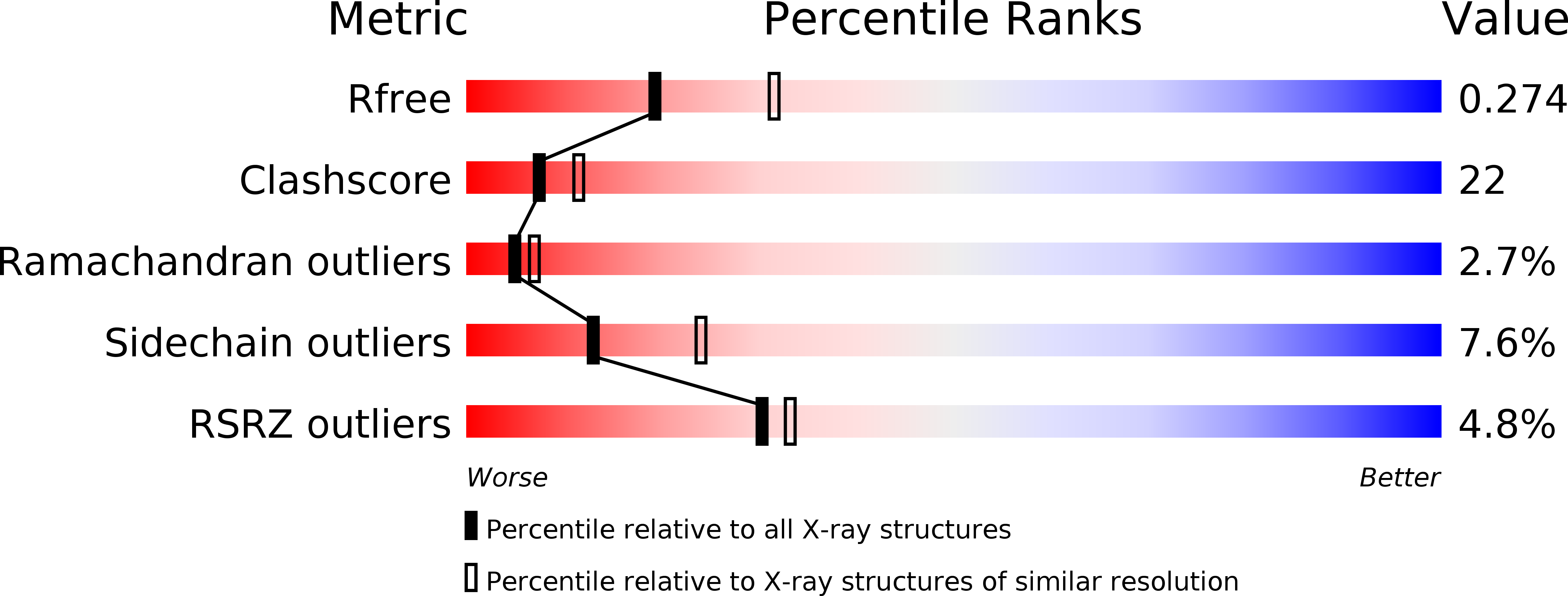
Resolution:
2.52 Å
R-Value Free:
0.27
R-Value Work:
0.22
R-Value Observed:
0.22
Space Group:
P 41 2 2
Macromolecular Entities
Polymer Type:polypeptide(L)
Molecule:PREDICTED: similar to ret finger protein-like 1
Chain IDs:A, B, C, D
Chain Length:201
Number of Molecules:4
Biological Source:Homo sapiens
Modified Residue
Compound ID Chain ID Parent Comp ID Details 2D Image
MSE A MET SELENOMETHIONINE
Primary Citation
Structure of the PRYSPRY-domain: Implications for autoinflammatory diseases
Febs Lett. 580 99 106 (2006)
PMID: 16364311 DOI: 10.1016/j.febslet.2005.11.076

Abstact

We determined the first structure of PRYSPRY, a domain found in over 500 different proteins, involved in innate immune signaling, cytokine signaling suppression, development, cell growth and retroviral restriction. The fold encompasses a 7-stranded and a 6-stranded antiparallel beta-sheet, arranged in a beta-sandwich. In the crystal, PRYSPRY forms a dimer where the C-terminus of an acceptor molecule binds to the concave surface of a donor molecule, which represents a putative interaction site. Mutations in the PRYSPRY domains of Pyrin, which are responsible for familial Mediterranean fever, map on the putative PRYSPRY interaction site.

Legend

Protein

Chemical

Disease

Primary Citation of related structures