2FBD image
Deposition Date 2005-12-09
Release Date 2006-12-12
Last Version Date 2024-10-30
Entry Detail
PDB ID:
2FBD
Keywords:
Title:
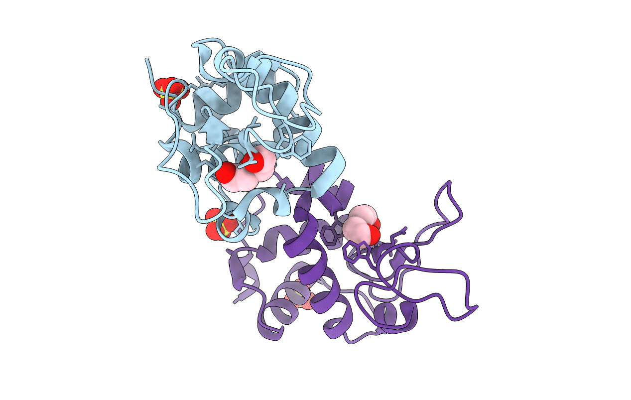
The crystallographic structure of the digestive lysozyme 1 from Musca domestica at 1.90 Ang.
Biological Source:
Source Organism:
Musca domestica (Taxon ID: 7370)
Host Organism:
Method Details:
Experimental Method:
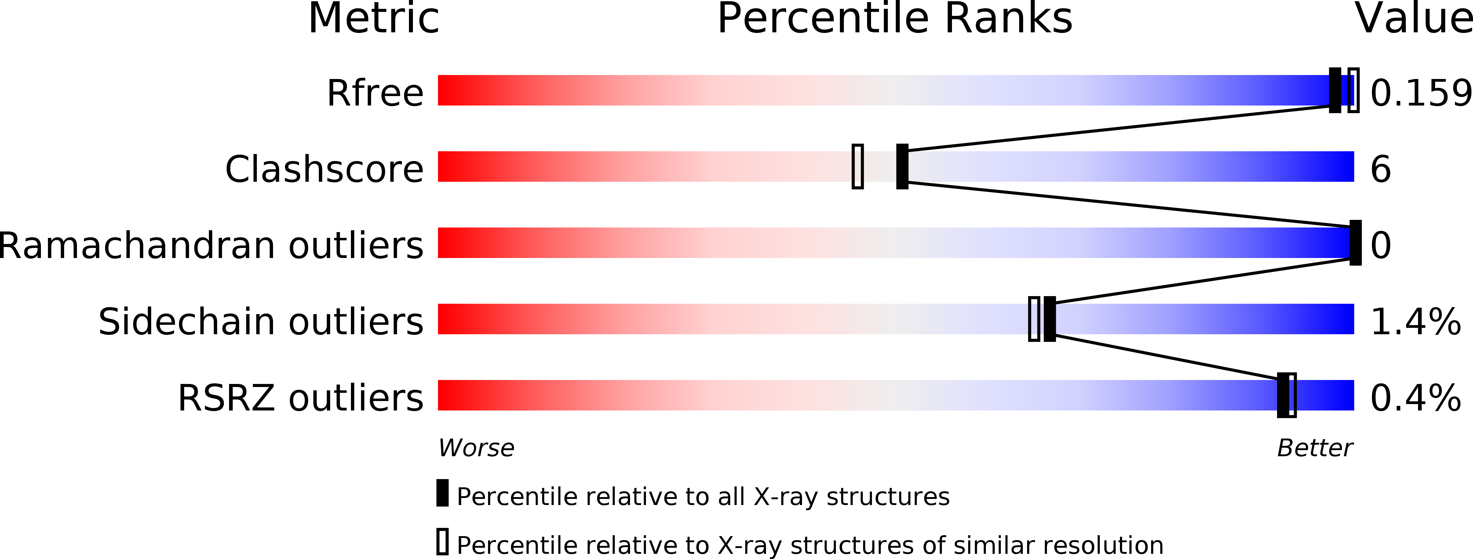
Resolution:
1.90 Å
R-Value Free:
0.19
R-Value Work:
0.15
R-Value Observed:
0.15
Space Group:
P 1 21 1
Macromolecular Entities
Polymer Type:polypeptide(L)
Molecule:Lysozyme 1
Chain IDs:A, B
Chain Length:122
Number of Molecules:2
Biological Source:Musca domestica
Primary Citation
Crystallization, data collection and phasing of two digestive lysozymes from Musca domestica.
Acta Crystallogr.,Sect.F 62 750 752 (2006)
PMID: 16880547 DOI: 10.1107/S1744309106024201

Abstact

Lysozymes are mostly known for their defensive role against bacteria, but in several animals lysozymes have a digestive function. Here, the initial crystallographic characterization of two digestive lysozymes from Musca domestica are presented. The proteins were crystallized using the sitting-drop vapour-diffusion method in the presence of ammonium sulfate or PEG/2-propanol as the precipitant. X-ray diffraction data were collected to a maximum resolution of 1.9 angstroms using synchrotron radiation. The lysozyme 1 and 2 crystals belong to the monoclinic space group P2(1) (unit-cell parameters a = 36.52, b = 79.44, c = 45.20 angstroms, beta = 102.97 degrees) and the orthorhombic space group P2(1)2(1)2 (unit-cell parameters a = 73.90, b = 96.40, c = 33.27 angstroms), respectively. The crystal structures were solved by molecular replacement and structure refinement is in progress.

Legend

Protein

Chemical

Disease

Primary Citation of related structures