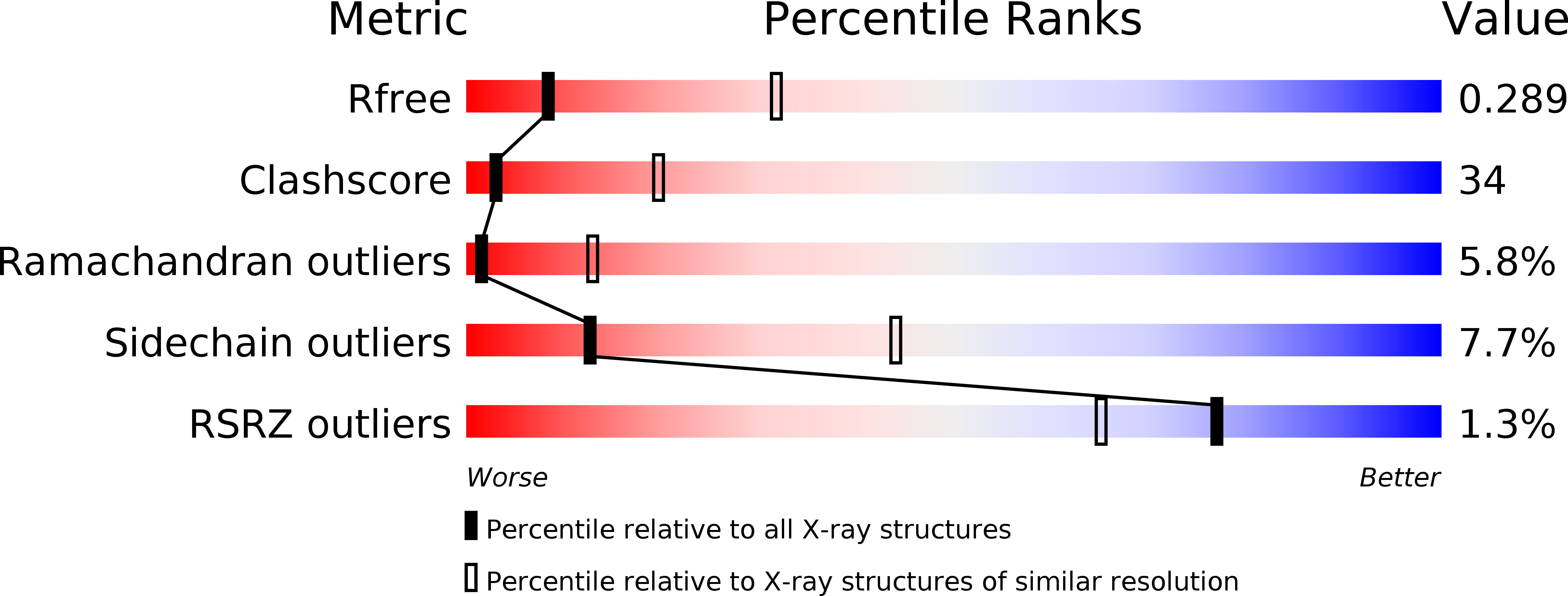
2F9Y image
Deposition Date 2005-12-06
Release Date 2006-12-12
Last Version Date 2024-10-16
Entry Detail
PDB ID:
2F9Y
Keywords:
Title:
The Crystal Structure of The Carboxyltransferase Subunit of ACC from Escherichia coli
Biological Source:
Source Organism:
Escherichia coli (Taxon ID: 562)
Host Organism:
Method Details:
Experimental Method:
Resolution:
3.20 Å
R-Value Free:
0.26
R-Value Work:
0.25
R-Value Observed:
0.25
Space Group:
P 65 2 2
Macromolecular Entities
Polymer Type:polypeptide(L)
Molecule:Acetyl-CoA carboxylase, Carboxyltransferase alpha chain
Chain IDs:A
Chain Length:339
Number of Molecules:1
Biological Source:Escherichia coli
Polymer Type:polypeptide(L)
Molecule:Acetyl-coenzyme A carboxylase carboxyl transferase subunit beta
Chain IDs:B
Chain Length:304
Number of Molecules:1
Biological Source:Escherichia coli
Ligand Molecules
Primary Citation
The Structure of the Carboxyltransferase Component of Acetyl-CoA Carboxylase Reveals a Zinc-Binding Motif Unique to the Bacterial Enzyme(,).
Biochemistry 45 1712 1722 (2006)
PMID: 16460018 DOI: 10.1021/bi0520479

Abstact

Acetyl-coA carboxylase (ACC) is a central metabolic enzyme that catalyzes the committed step in fatty acid biosynthesis: biotin-dependent conversion of acetyl-coA to malonyl-coA. The bacterial carboxyltransferase (CT) subunit of ACC is a target for the design of novel therapeutics that combat severe, hospital-acquired infections resistant to the established classes of frontline antimicrobials. Here, we present the structures of the bacterial CT subunits from two prevalent nosocomial pathogens, Staphylococcus aureus and Escherichia coli, at a resolution of 2.0 and 3.0 A, respectively. Both structures reveal a small, independent zinc-binding domain that lacks a complement in the primary sequence or structure of the eukaryotic homologue.

Legend

Protein

Chemical

Disease

Primary Citation of related structures